
Fig. 7
- ID
- ZDB-IMAGE-180302-27
- Genes
- Publication
- Seritrakul et al., 2017 - Tet-mediated DNA hydroxymethylation regulates retinal neurogenesis by modulating cell-extrinsic signaling pathways
- All Figures
- Figures for Seritrakul et al., 2017
|
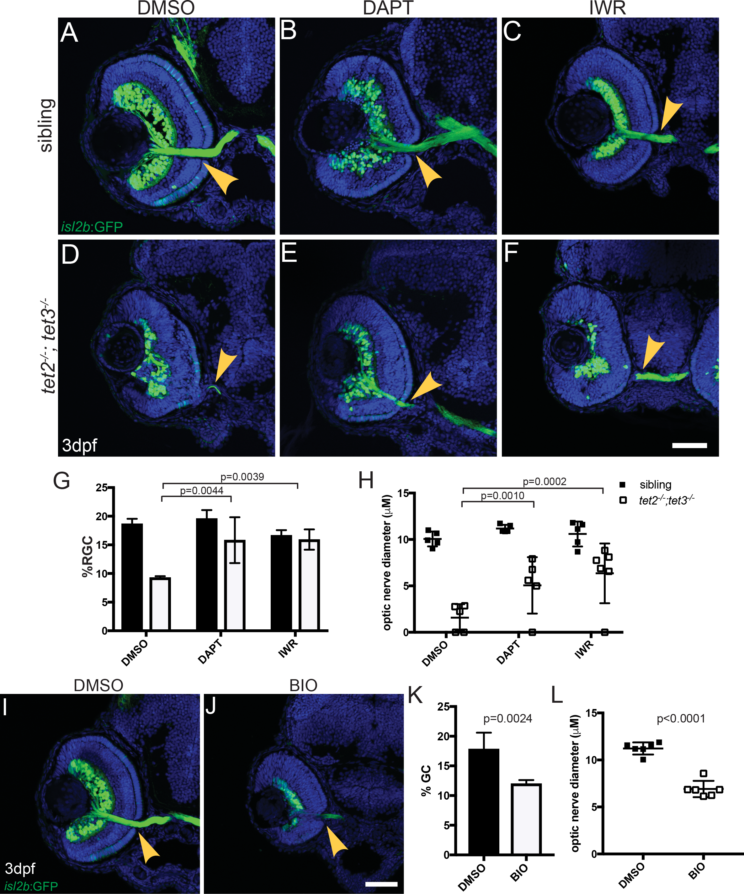
Fig. 7
tet2-/-;tet3-/- mutant and sibling embryos carrying isl2b:GFP were exposed to 50μM DAPT, 5μM IWR-1-endo, or 1% DMSO, during neurogenesis (24hpf to 72hpf) and analyzed for isl2b:GFP+ RGCs and axons. (A-C, G-H) No significant increases in optic nerve diameter or percentage of isl2b:GFP+ RGC per total cells (%RGC) were detected in sibling embryos treated with either 1% DMSO (vehicle), DAPT (Notch inhibitor), or IWR (Wnt inhibitor). (E,G,H) tet2-/-;tet3-/- mutants treated with 50μM DAPT showed a significant increase in both the percentage of isl2b:GFP+ RGCs per retina and optic nerve diameter, when compared to DMSO-treated controls. (F-H) tet2-/-;tet3-/- embryos treated with 5μM IWR also showed a significant increase in the percentage of isl2b:GFP+ RGCs per retina and optic nerve diameter. (I-L) Wnt signaling was upregulated by exposing wild-type embryos from 24hpf to 72hpf with 2μM BIO, a GSK3β inhibitor. (I,J) BIO-treated wild-type embryos showed reduced lamination, (K) a decreased percentage of isl2b:GFP+ RGCs per retina, and (L) decreased optic nerve diameter relative to DMSO-treated controls, (p = 0.00236 and p<0.0001). All error bars = ± 1 S.D.; n = 5 embryos (A-H) and n = 6 embryos (I-L) per condition analyzed; P-values calculated using two-way ANOVA with multiple comparisons (for G-H) and two-tailed, unpaired t-test (for K-L). Scale bar = 50μm.