
Fig. 2
- ID
- ZDB-IMAGE-180208-17
- Publication
- Dudczig et al., 2017 - Developmental and adult characterization of secretagogin expressing amacrine cells in zebrafish retina
- All Figures
- Figures for Dudczig et al., 2017
|
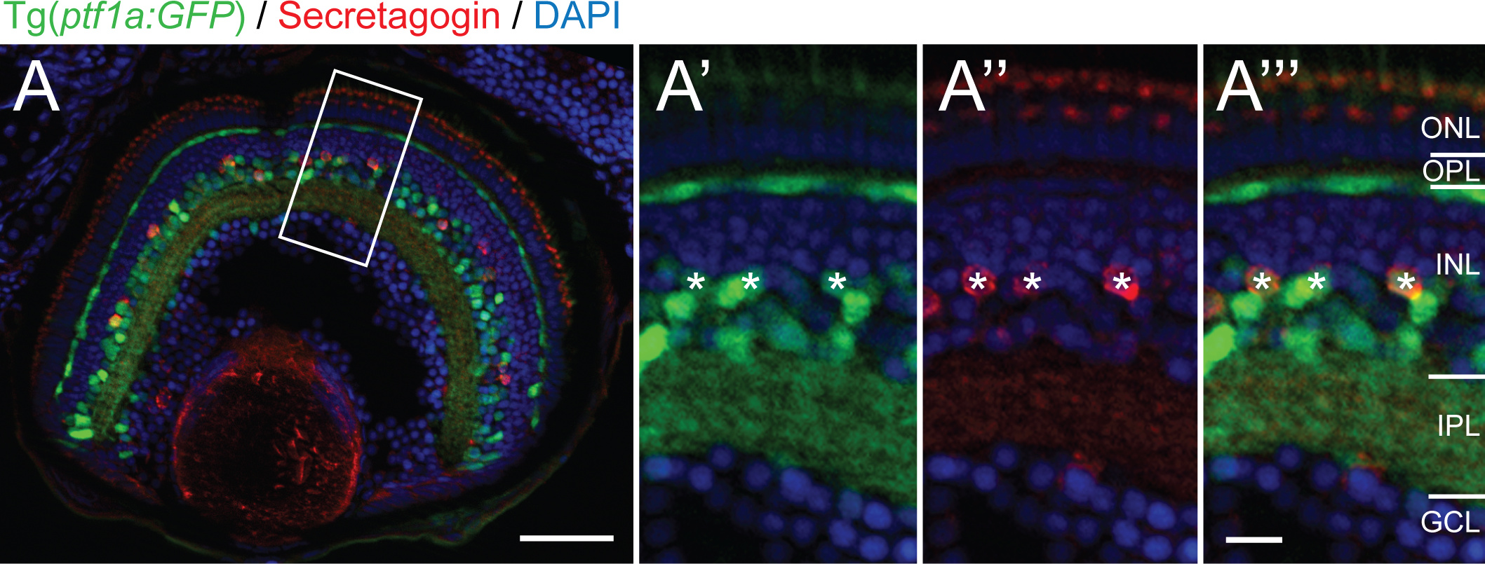
Fig. 2
Secretagogin positive cells co-label with the Ptf1a:GFP amacrine marker in the inner nuclear layer.
Micrograph at 5 days postfertilization showing secretagogin immunostained Tg(ptf1a:GFP) zebrafish retinas. Higher magnification of boxed view of boxed inset shows co-localization (asterisks) of SCGN+ (red) and Ptf1a:GFP+ (green) within individual cells marked in the green (A'), red (A'') and double (A''') channels.). ONL: outer nuclear layer; OPL: outer plexiform layer; INL: inner nuclear layer; IPL: inner plexiform layer; GCL: ganglion cell layer. Scale bar (A) is 50 μm, scale bar (A''') for A'–A''' is 10 μm.