
Fig. S9
- ID
- ZDB-IMAGE-180206-7
- Publication
- Singh et al., 2017 - Different developmental histories of beta-cells generate functional and proliferative heterogeneity during islet growth
- All Figures
- Figures for Singh et al., 2017
|
Fig. S9
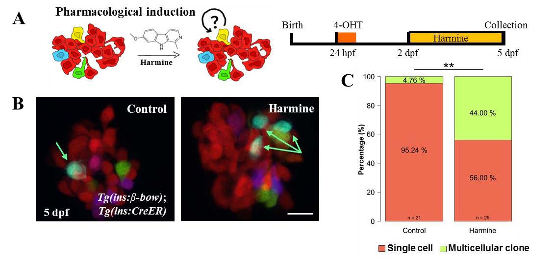
Small molecule based reversal of DBCs quiescence using harmine. (A) Cartoon (left) and schematic (right) for the pharmacological stimulation of proliferation. Dorsal bud-derived beta-cells (DBCs) were labeled by incubating beta-bow animals with 4-OHT at 24 hpf. Subsequently, the animals were incubated from 2–5 dpf with Harmine or DMSO. (B) Maximum intensity projections of the primary islets from control and harmine-treated beta-bow animals. Arrows indicate trichromatic cells. (C) Quantification of the proportion of trichromatic beta-cells that remain as single cells or form multicellular clones (≥ 2 cells/clone) in the control and laser-ablated islets. Harmine-treatment increased the percentage of multicellular clones compared to DMSO (Fisher’s exact test, ** p ≤ 0.01). Scale bars, 10 μm.