
IMAGE
Fig. 4
- ID
- ZDB-IMAGE-180124-17
- Publication
- Tokunaga et al., 2017 - Comprehensive validation of T- and B-cell deficiency in rag1-null zebrafish: Implication for the robust innate defense mechanisms of teleosts
- All Figures
- Figures for Tokunaga et al., 2017
Image
|
Figure Caption
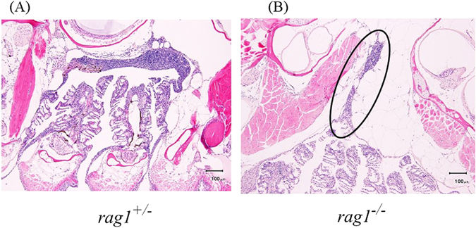
Fig. 4
Histological observation of the hematoxylin–eosin-stained thymus in rag1 +/− (left) and rag1 −/− fish (right). The thymus in rag1 −/− fish is representative of five independent samples. The thymus in rag1 +/− fish is representative of three independent samples. A thymus was not found in two rag1 +/− fish. The circle shows atrophic lymphocyte clump.
Figure Data
Acknowledgments
This image is the copyrighted work of the attributed author or publisher, and
ZFIN has permission only to display this image to its users.
Additional permissions should be obtained from the applicable author or publisher of the image.
Full text @ Sci. Rep.