
|
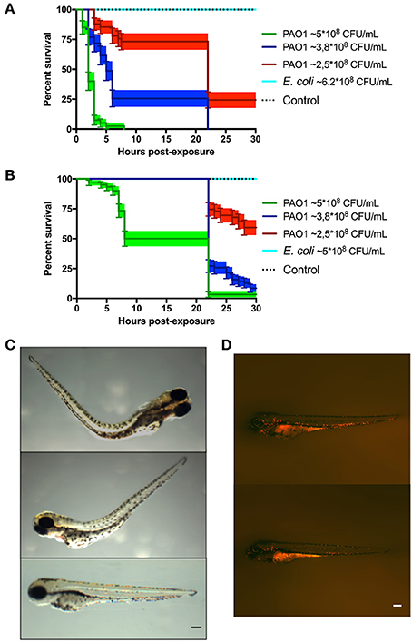
Fig. 1
Zebrafish larvae exposed to P. aeruginosa by static immersion. Larvae were immersed at 72 HPF in P. aeruginosa PAO1, E. coli DH5α or in sterile E3 medium as control. (A,B) Survival curve of 72 HPF larvae immersed in different suspensions P. aeruginosa PAO1 (green, blue and red) or in a suspension of E. coli DH5α (light blue line) or in sterile E3 medium (black dotted line). In (A) the bacteria were grown in PGS (↓Pi) medium. In (B) the bacteria were grown in PGS (↑Pi) medium. (C) Larvae immersed with ~5 × 10⁸ CFU/mL of P. aeruginosa PAO1 grown in PGS (↓Pi) medium (upper and second picture) or in sterile E3 medium (bottom picture) at 3 hpe. (D) Tg(BACmpo:mCherry) larvae immersed at 72 HPF in ~2.5 × 10⁸ CFC/mL of P. aeruginosa PA01 grown in (↑Pi) medium (top) or in sterile E3 medium (bottom) at 22 hpe. Scale (A,B): 100 μm.