
|
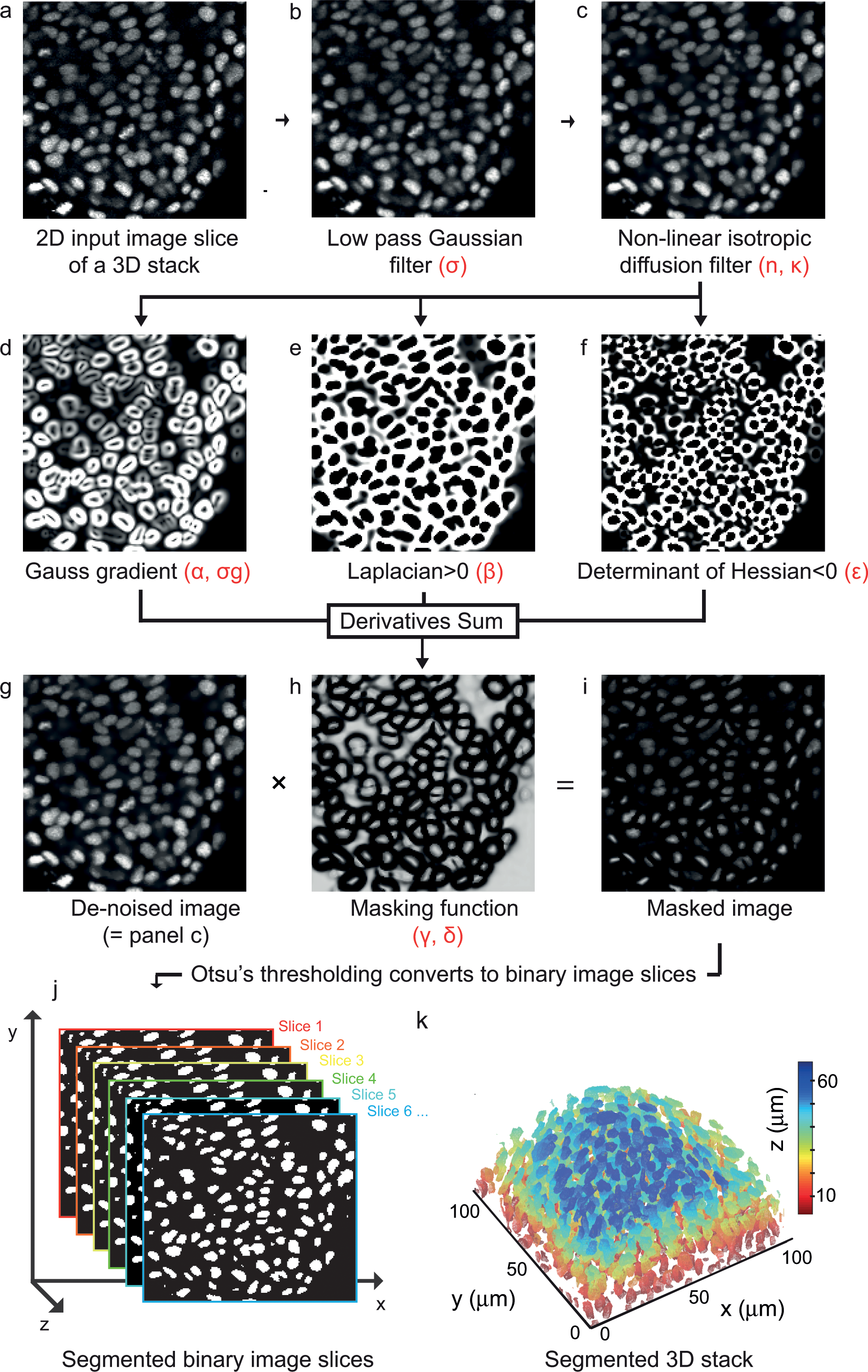
Fig. 1
Derivatives sum algorithm for 3D nuclear segmentation.
(a) 2D 8-bit gray scale image slice of a 3D stack of dimension 167 × 172 × 39 voxels from the posterior PSM of an 18-somite stage zebrafish embryo. (b) De-noised image after Gaussian blur (σ = 0.5, window size = 5×5 squared pixels). (c) Image smoothened by a non-linear isotropic diffusion filter (κ = 10, n = 4). (d) Magnitude of Gauss gradient (σg = 1.5), (e) Laplacian where positive and (f) Determinant of Hessian where negative, we show the absolute value. (g) De-noised image shown in (c). (h) A tangent hyperbolic masking function (α = β = ε = γ = δ = 1). (i) Masked image obtained by the pair-wise product between the de-noised image in (g) and the masking function in (h). (j) Slices of binary images obtained by Otsu’s thresholding method. (k) Surface rendered 3D binary objects colored with respect to their position along the z-direction. All tunable parameters are highlighted by Greek glyphs in red. See S1 Text for more details.