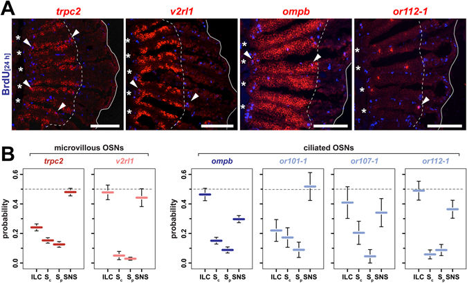
Fig. 5
Functional equivalence and bias in OSN neurogenesis at the ILC and SNS. (A) In situ-hybridization (red) for markers of distinct OSN subtypes and immunohistochemistry against BrdU (blue) at 8d (trpc2, ompb) and 4d (v2rl1, or112-1) following 24 h BrdU incubation. The trpc2 gene is expressed by all microvillous OSNs, a larger subset of which also expresses the vomeronasal receptor v2rl1. Ciliated OSNs label positive for ompb and express specific olfactory receptor genes, such as or112-1. For each gene, double-positive cells (arrowheads) can be found at the interlamellar curves (asterisks) and the sensory/nonsensory border (dotted line). The solid line demarcates the outline of the section of which only one half is shown. Scale bars: 100 µm. (B) Quantification of OSN subtype neurogenesis at different proliferation sites. The sensory OE was subdivided into four equal segments (ILC: interlamellar curves, Sc: sensory central, Sp: sensory peripheral, SNS: sensory/nonsensory border) and the occurrence of marker/BrdU double-positive OSNs was scored for each segment for the general microvillous marker trpc2 (red), the vomeronasal-like receptor v2rl1 (light red), the ciliated OSN marker ompb (blue) and the three classical OR genes or101-1, or107-1, and or112-1 (light blue) expressed by ciliated OSNs. A subtype-specific bias exists for neurogenesis at the ILC and SNS. Data represent the mean ± SEM; trpc2: 607 cells from 5 OE; v2rl1: 203 cells from 4 OE; ompb: 563 cells from 2 OE; or101-1: 35 cells from 6 OE; or107: 25 cells from 7 OE; and or112-1: 76 cells from 8 OE.

