
|
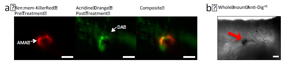
Fig. S1
Further staining to confirm cell death. (a) 3 dpf ren:mem-KillerRed fish were incubated in acridine orange (2 ng/ml) for 30 mins in the dark, followed by two 5 mins washes and immediately imaged live on the SPIM. Treatment with the light-sheet was performed (1 hr, 4 mW, depth-stack every 10s) and the acridine orange fluorescence signal was collected post-treatment and a composite image was created. Treated KillerRed+ cells can be seen to take up acridine orange readily. (b) A whole mount in situ protocol was performed with an anti-Dig+ antibody, visualised using the colourant Nitro Blue Tetrazolium using the brightfield camera on the SPIM. Dig+ cells are dark spots, as indicated with the red arrow. Scale bars represent 30 μm.