
|
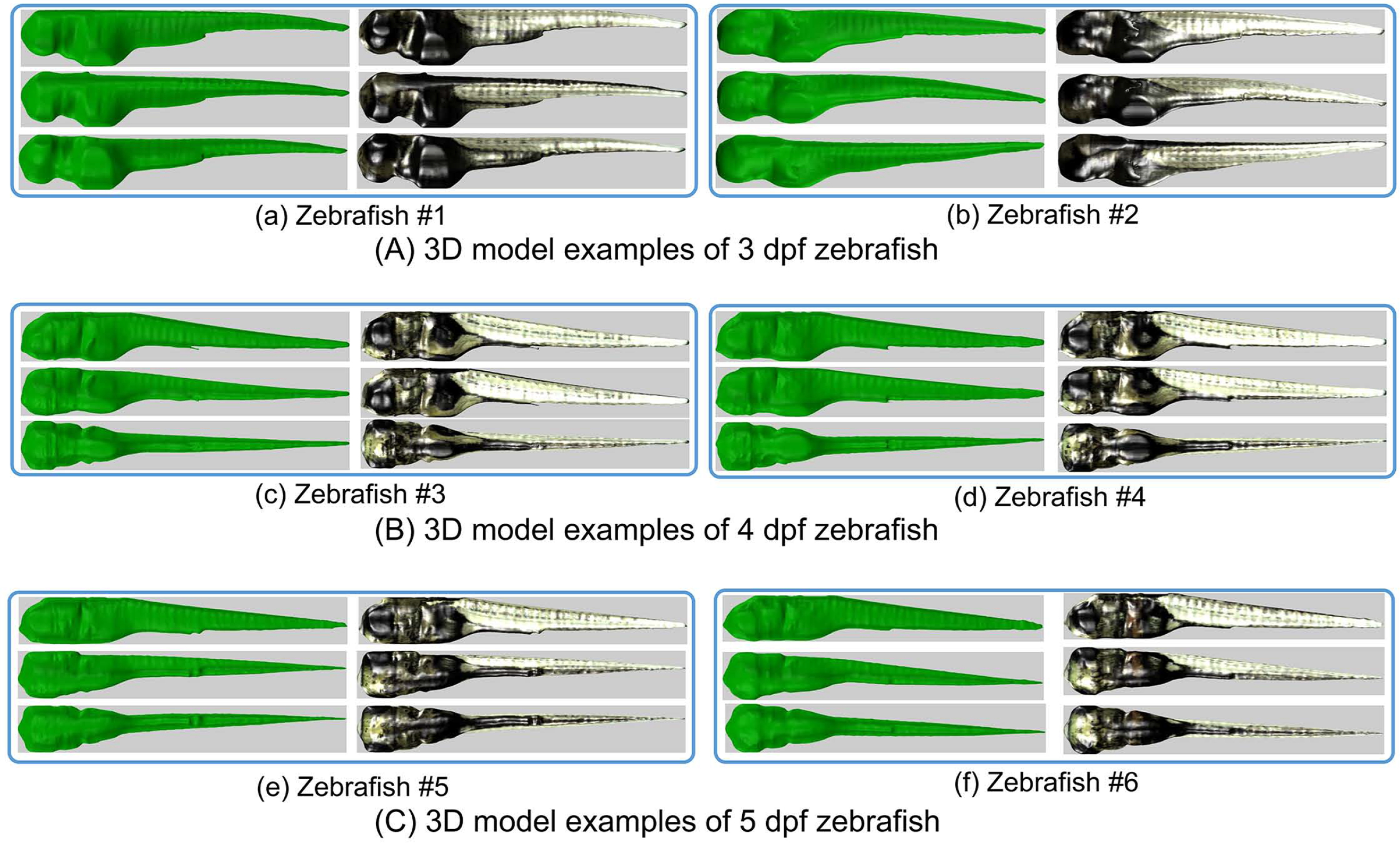
Fig. 10
Visualization of 3D models of 3 zebrafish larval stages (3 dpf, 4 dpf and 5 dpf). Each box represents a reconstructed 3D model for one specific zebrafish larvae visualized from three different viewpoints. The 3D volumetric representations are shown in green on the left side in each box. The models with texture-mapping are in the right side of each box. (A) 3D models of two selected 3 dpf zebrafish larvae. (B) 3D models of two selected 4 dpf zebrafish larvae. (C) 3D models of two selected 5 dpf zebrafish larvae. Variation in size and shape between stages and within stages (interclass and intraclass) can be appreciated from the visualizations. A remarkable intraclass discrimination originates from the size and color of the yolk. In addition, animations of the 3D zebrafish models are available at: http://bio-imaging.liacs.nl/galleries/VAST-3Dimg/.