
|
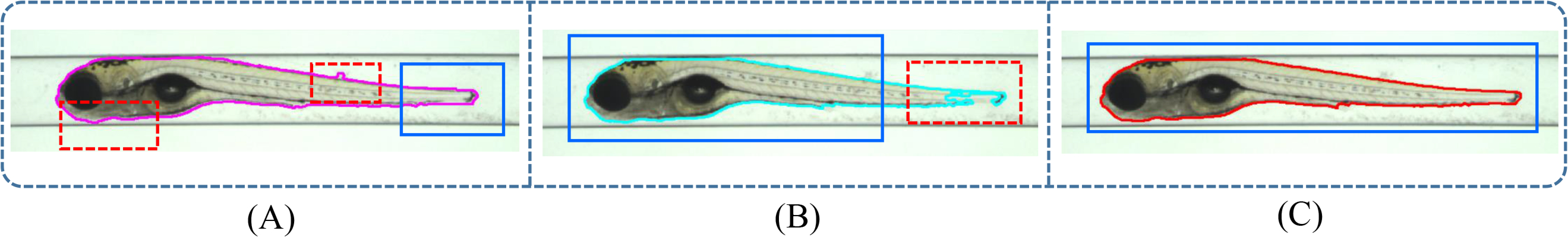
Fig. 3
Results of various segmentation methods for zebrafish in VAST. The blue bounding box indicates an accurate part of the segmentation; red bounding box indicates an inaccurate part of the segmentation. (A) The segmentation obtained by the mean shift algorithm. This method produces a whole shape representation of the zebrafish but fails in sensitivity to the edges. (B) The segmentation obtained by the improved level set method. This method fails to detect the transparent regions as found in zebrafish tail area. (C) An accurate segmentation is obtained from a hybrid approach including the previous two methods with additional refinements.