
Fig. 4
- ID
- ZDB-IMAGE-180108-3
- Genes
- Publication
- Figueroa et al., 2017 - Reprimo tissue-specific expression pattern is conserved between zebrafish and human
- All Figures
- Figures for Figueroa et al., 2017
|
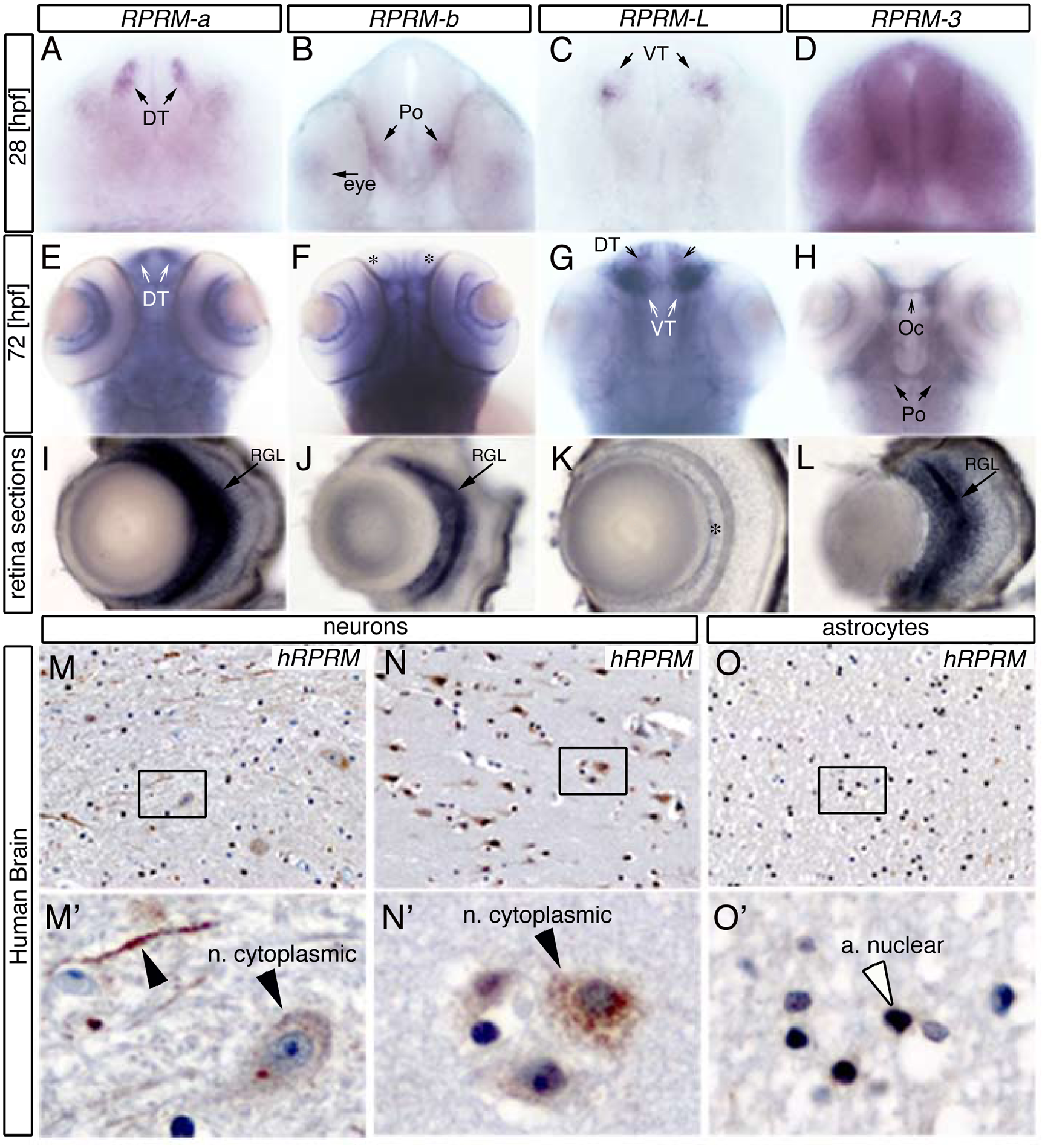
Fig. 4
RPRM expression patterns are conserved between zebrafish and human brain.
(A-D) RPRM expression patterns were examined using whole-mount in situ hybridization in wild-type embryos at 28 and (E-H) 72 hours post-fertilization [hpf]. (A-D) Frontal views of the embryo heads. (E-H) Dorsal views of the embryo heads. (I-L) Retina cross-sections. (A-C) At 28 hpf rprma, rprmb and rprml transcripts are expressed in neuronal populations such as dorsal thalamus (DT), preoptic region (Po) and ventral thalamus, respectively (black arrows). (D) rprm3 is ubiquitously expressed throughout the brain. (E-G) At 72hpf rprma and rprml are expressed in the DT and the VT, respectively (white arrows), while rprmb is not expressed in those regions (asterisks). (H) At the same developmental stage, rprm3 mRNA is expressed in the Po and the optic chiasma (Oc). (I-L) Cross sections of the retina. (I, J, L) rprma, rprmb and rprm3 expression are restricted in the retina to the retinal ganglion cell layer (RGL, black arrow). (K) In contrast, rprml transcript expression is absent in the RGL (asterisk). (M, M’, N, N’) IHC staining for RPRM of white and grey matter sections from adult human samples (400x; inset magnifications 600x). (M-N) RPRM protein is expressed in the cytoplasm and axons of neurons (black arrowheads). (O-O’) RPRM protein is expressed in the nuclei of astrocytes.