
|
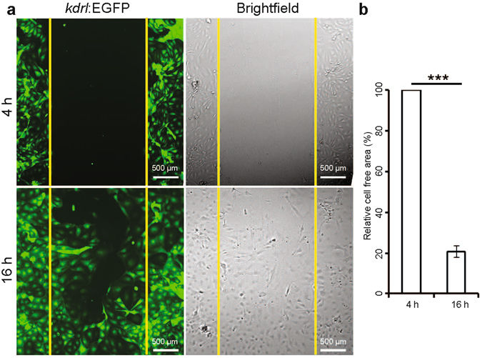
Fig. 6
Scratch/wound-healing assay on cardiac endothelial cells. Assay was performed on isolated cardiac endothelial cells from adult Tg(kdrl:EGFP) zebrafish ventricles. (a) Fluorescence and brightfield images at 4 and 16 h after scratching. (b) Quantification of the initial cell free area covered by endothelial cell migration in 12 h. Cell free area at 4 h after scratch was considered as 100% (n = 3, mean ± SEM). One way ANOVA followed by Bonferroni’s post-hoc test (GraphPad Prism) was performed to evaluate statistical significance of differences. P < 0.05 was considered statistically significant. *** corresponds to P < 0.001.