
Fig. 1
- ID
- ZDB-IMAGE-180108-11
- Publication
- Bremer et al., 2017 - A small molecule screen identifies in vivo modulators of peripheral nerve regeneration in zebrafish
- All Figures
- Figures for Bremer et al., 2017
|
Fig. 1
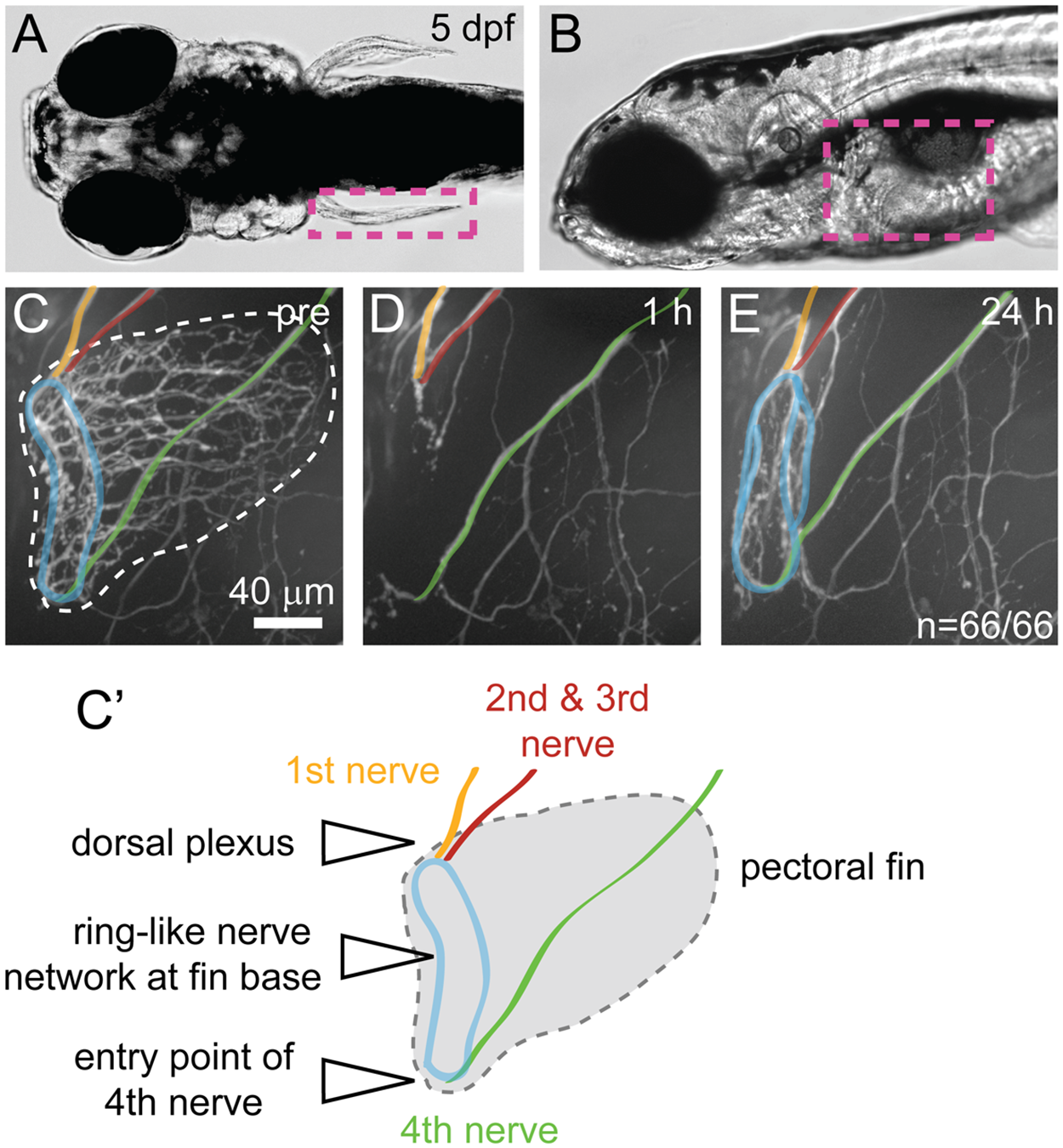
Pectoral fin innervation and nerve regrowth after fin removal.
(A-B) Dorsal (A) and side view (B) of a larval zebrafish at 5 dpf showing the location of the pectoral fin (magenta dashed box). (C, C') Pectoral fin innervation in a Tg(mnx1:GFP) transgenic larva at 5 dpf (C) and a schematic (C'). The first segmental nerve (orange) forms the dorsal plexus together with the joint nerves 2 and 3 (red). The fourth nerve (green) travels along the larval body wall, before it enters the pectoral fin ventrally. These nerves contribute to the ring-like network at the fin base (blue). (D-E) Fin region of Tg(mnx1:GFP) transgenic larvae 1 hour (D) and 24 hours (E) after fin removal. One hour after the pectoral fin has been removed using tweezers, GFP positive axons at the fin base are no longer detectable, leaving behind the stumps of nerve 1–3 (orange, red) at the dorsal plexus and at the 4th nerve entry point (green, D). Twenty-four hours after fin removal, GFP positive axons at the fin base have regrown robustly, reforming the ring-like nerve network at the fin base (blue) in 100% of the 66 control fish tested (untreated and DMSO treated, E).