
IMAGE
Fig. 2
- ID
- ZDB-IMAGE-180105-45
- Publication
- Smith et al., 2017 - TNFa/TNFR2 signaling is required for glial ensheathment at the dorsal root entry zone
- All Figures
- Figures for Smith et al., 2017
Image
|
Figure Caption
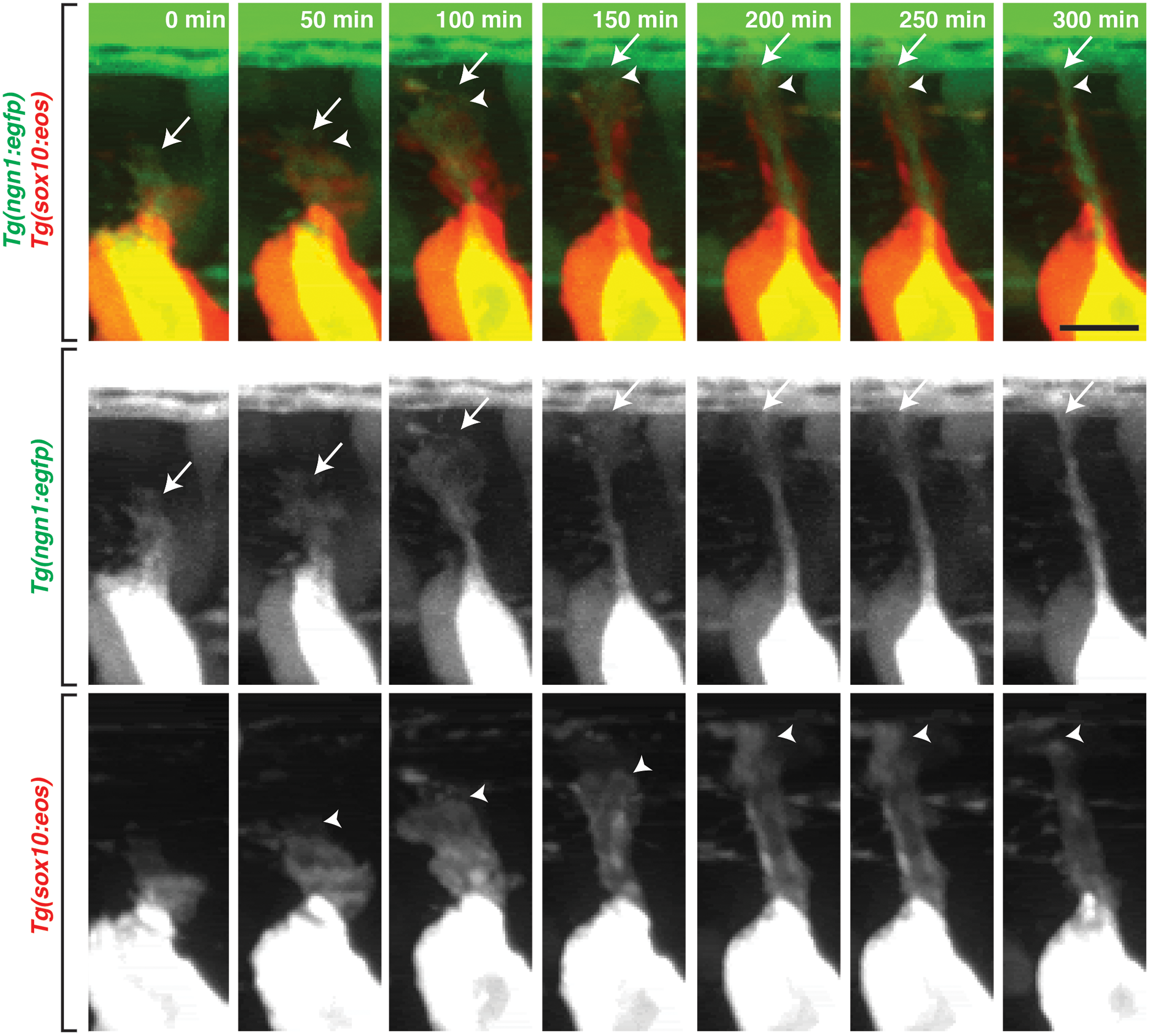
Fig. 2
DRG glia navigate with pioneer axons.
Images from a 24 hour time-lapse movie starting at 48 hpf in a Tg(sox10:eos);Tg(ngn1:egfp) embryo that was exposed to UV light to photoconvert Eos in the whole embryo. These frames show the pioneer axon (green) and associated glia (red) migrate together to the DREZ. Below are individual channels for GFP and Eos. Arrows denote the pioneer axon growth cone and arrowheads denote location of the DRG glia. Scale bar, 25 μm.
Acknowledgments
This image is the copyrighted work of the attributed author or publisher, and
ZFIN has permission only to display this image to its users.
Additional permissions should be obtained from the applicable author or publisher of the image.
Full text @ PLoS Genet.