
Image
|
Figure Caption
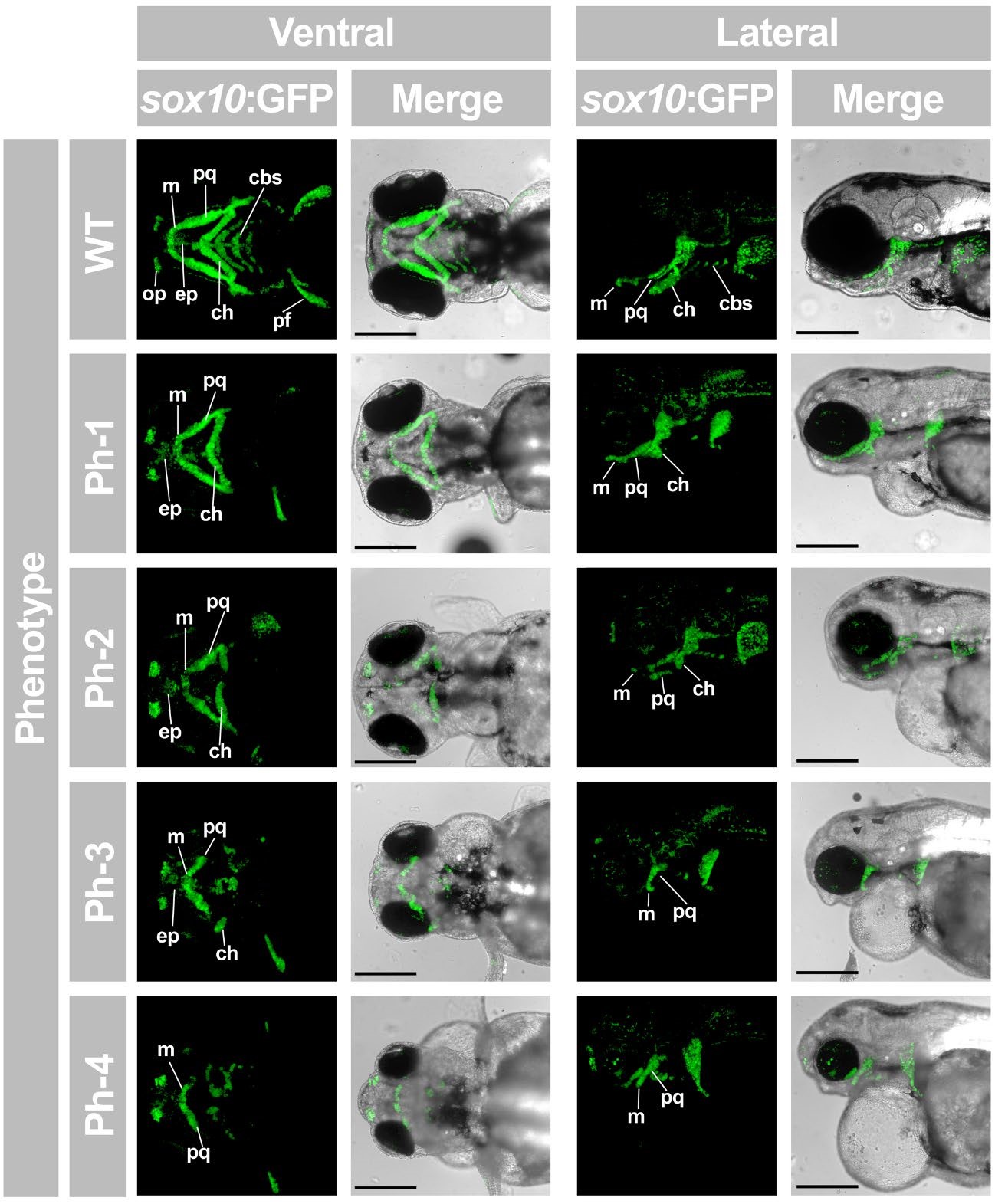
Fig. S10
Characterization of jaw cartilague maldevelopment of Gpatch3ATG morphants at 96 hpf.
Reporter Tg(sox10:GFP) zebrafish embryos were microinjected with 2.0 ng of the gpatch3ATG MO. Merge of the maximum intensity projection of fluorescent signals (sox10:GFP) and transmitted light micrographs is also shown. cbs, ceratobranchials; ch, ceratohyal, m, Meckel’s cartilage; ep, ethmoid plate; pq, palatoquadrate; op, olfactory pit; pf, pectoral fin. Scale bars represent 200 μm.
Acknowledgments
This image is the copyrighted work of the attributed author or publisher, and
ZFIN has permission only to display this image to its users.
Additional permissions should be obtained from the applicable author or publisher of the image.
Full text @ Sci. Rep.