Fig. S1
Degeneration of cone and rod photoreceptors at 4 wpf in the gosh mutant
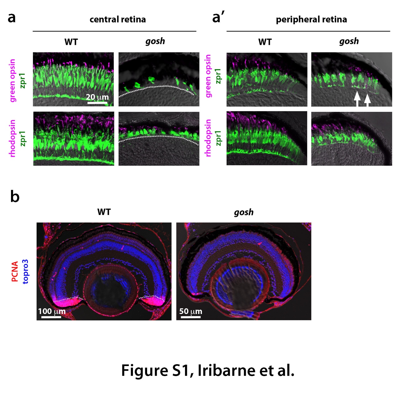
(a) Labeling of wild-type and gosh mutant retinas at 4 wpf with green opsin and zpr1 antibodies, or rhodopsin and zpr1 antibodies. In wild-type central retina, the ONL is thicker than at 7 dpf to segregate rod and cone nuclear layers. Its cone shape is highly elongated. Opsin and rhodopsin are localized in the OS. In gosh mutant central retina, the ONL is very thin, and cones are fewer in number and abnormal in shape. (a') In wild-type peripheral retina, new rod and cone photoreceptors are generated and express rhodopsin and opsin, respectively. In gosh mutant peripheral retina, cones become columnar although their thickness is still less than in wild type retina. Green opsin and rhodopsin are detected; however, green opsin is mislocalized through the cell body (arrows). A dotted line indicates the boundary between photoreceptor cells and INL. (b) Labeling of wild-type and gosh mutant retinas of 2 wpf with PCNA antibody (red). Nuclei are counter-stained with TOPRO3 (blue). In wild-type retina, most of the CMZ express PCNA, indicating cell proliferation of retinal stem and progenitor cells (dotted line). In wildtype retina, PCNA positive cells are observed just beneath the ONL and in the INL, which correspond to rod progenitors and Müller cell-derived regenerating progenitors, respectively. In contrast, PCNA expression is reduced in the gosh mutant CMZ, in the number of rod progenitors and Müller cell-derived regenerating progenitors, suggesting that retinal regeneration is not active in the gosh mutant at 2 wpf.

