
Fig. 5 S1
- ID
- ZDB-IMAGE-180103-22
- Publication
- Sidhaye et al., 2017 - Concerted action of neuroepithelial basal shrinkage and active epithelial migration ensures efficient optic cup morphogenesis
- All Figures
- Figures for Sidhaye et al., 2017
|
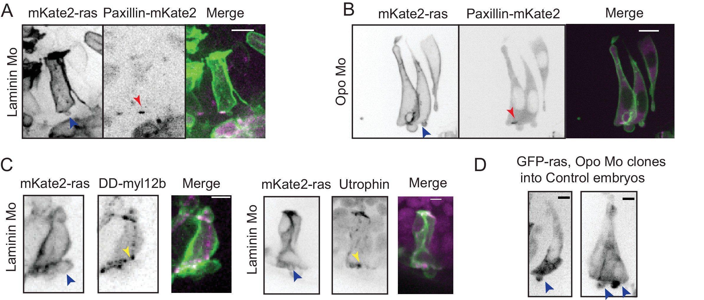
Fig. 5 S1
Effect of perturbed cell-ECM attachment on the rim cells.
(A) Confocal scan of rim cell in laminin morphant embryos with mosaic expression of mKate2-ras and paxillin-mKate2. Blue arrowhead points at the bleb. Red arrowhead marks at the stable paxillin localization. Frames from Video 15. N = 3. Scale bar = 10 µm. (B) Confocal scan of rim cell in opo morphant embryos with mosaic expression of mKate2-ras and paxillin-mKate2. Blue arrowhead points at the bleb. Red arrowhead marks at the transient paxillin localization. Frames from Video 16. N = 3. Scale bar = 10 µm. (C) Confocal scan of rim cells exhibiting blebs (blue arrowhead) in laminin morphants mosaically labeled by mKate2-ras with DD-myl12b-GFP (left) and GFP-UtrophinCH (right). Yellow arrowhead marks the localization of DD-myl12b-GFP outside and UtrophinCH inside the bleb. Scale bar = 2 µm. (D) Confocal scans of rim zone showing GFP-ras expressing opo morphant clone in control embryos. N = 4 out of 5 transplanted embryos. Blue arrow points at the basal blebs exhibited by the transplanted morphants cells. Scale bar = 5 µm.