
Fig. 2 S1
- ID
- ZDB-IMAGE-180103-14
- Publication
- Sidhaye et al., 2017 - Concerted action of neuroepithelial basal shrinkage and active epithelial migration ensures efficient optic cup morphogenesis
- All Figures
- Figures for Sidhaye et al., 2017
|
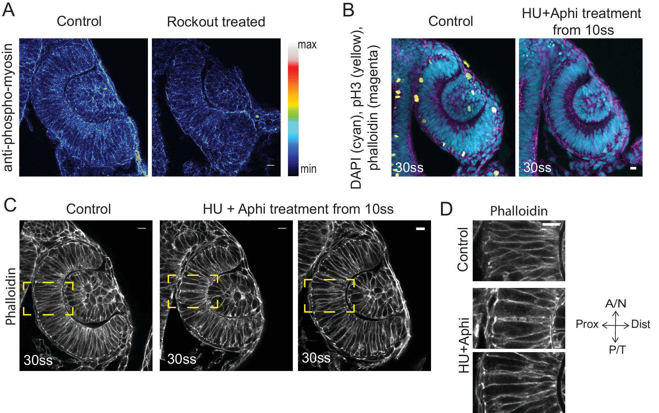
Fig. 2 S1
Effect of Rockout and HU+Aphi treatment on the RNE.
(A) Confocal scan of 30 ss RNE in control (left) and Rockout treated (right) embryo stained for phosphomyosin.Rockout treatment was performed for 7 h starting from 13–14 ss. Lookup table indicates the minimum and maximum intensity values. (B) Confocal scans of 30 ss RNE in control (left) and HU+ Aphi treated (right) embryos stained for DAPI (cyan), mitotic marker phosphohistone-3 (yellow) and phalloidin (magenta). (C) Confocal scans of 30 ss RNE in control (left) and HU+ Aphi-treated (middle and right) embryos stained with phalloidin. Areas marked by yellow box are shown in D. (D) Confocal scans of RNE cells (zoomed images of regions marked in C). Control (top), HU+Aphi treated (middle and bottom) stained with phalloidin. All scale bars = 10 µm.