
Image
|
Figure Caption
Fig. 4
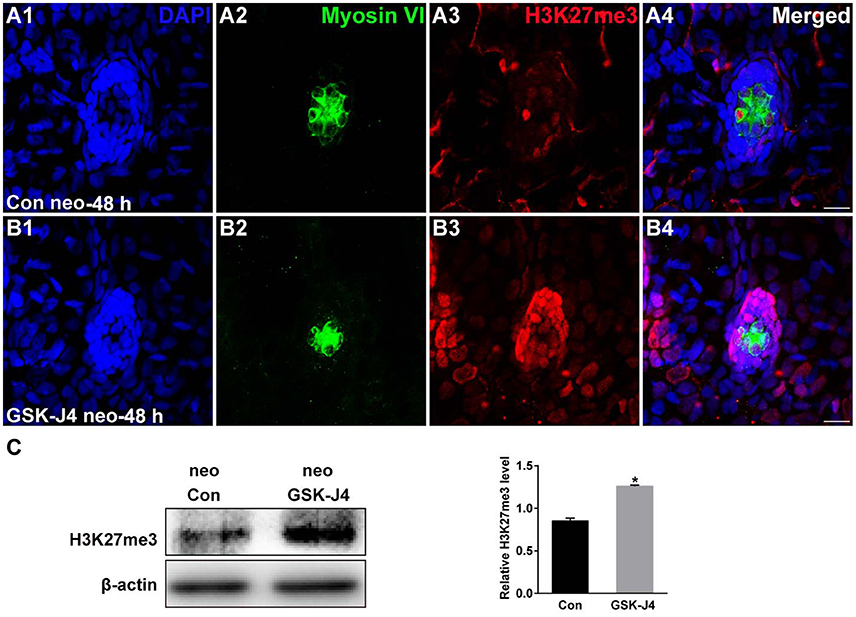
GSK-J4 increased the levels of H3K27me3. (A,B) Immunohistochemistry results showing H3K27me3 expression in neuromasts of DMSO-treated control (Con) larvae (n = 5 larvae) and of 10 μM GSK-J4-treated larvae (n = 6 larvae) for 48 h. Scale bar = 10 μm. (C) Western blot analysis was performed to evaluate the expressions of H3K27me3 and β-actin in whole larvae that were treated with either DMSO (neo Con; n = 6 larvae) or GSK-J4 (neo GSK-J4; n = 6 larvae) for 48 h. *p < 0.05.
Acknowledgments
This image is the copyrighted work of the attributed author or publisher, and
ZFIN has permission only to display this image to its users.
Additional permissions should be obtained from the applicable author or publisher of the image.
Full text @ Front. Mol. Neurosci.