
Image
|
Figure Caption
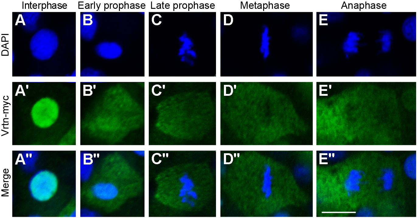
Fig. S13
Nuclear localisation of Vrtn during interphase of the cell cycle. Immuofluorescence staining of Vrtn-myc during different phases of the cell cycle in zebrafish embryos at 50% epiboly. (A-E) DAPI staining of nuclear DNA. (A'-E') Immunofluorescence staining of Vrtn-myc. Nuclear localisation is detected only during the interphase. (A''-E'') Merge of DAPI and Vrtn-myc staining. Scale bar: 10 μm.
Acknowledgments
This image is the copyrighted work of the attributed author or publisher, and
ZFIN has permission only to display this image to its users.
Additional permissions should be obtained from the applicable author or publisher of the image.
Full text @ Development