
|
Fig. S3
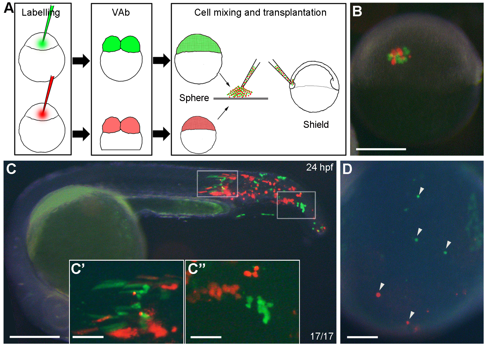
Transplanted cells from VAb@2cell embryos can differentiate into ventral tissues. (A) Diagram illustrating the procedure of cell transplantation to compare the distribution of descendent cells from intact (green) and VAb@2cell (red) embryos in unlabelled intact recipients. (B) A successfully transplanted embryo at shield stage. (C-C'') Comparable localisation of green and red cells in a chimeric embryo at 24 hpf, with higher magnifications of the boxed regions. (D) Transplanted cells from intact and VAb@2cell embryos can similarly differentiate into circulating blood cells (arrowheads) in the yolk sac circulation valley of a 24 hpf chimera. Scale bars: (B,C) 250 μm; (C'-D) 50 μm.