
Fig. 2
- ID
- ZDB-IMAGE-171205-7
- Publication
- Just et al., 2016 - Tbx20 Is an Essential Regulator of Embryonic Heart Growth in Zebrafish
- All Figures
- Figures for Just et al., 2016
|
Fig. 2
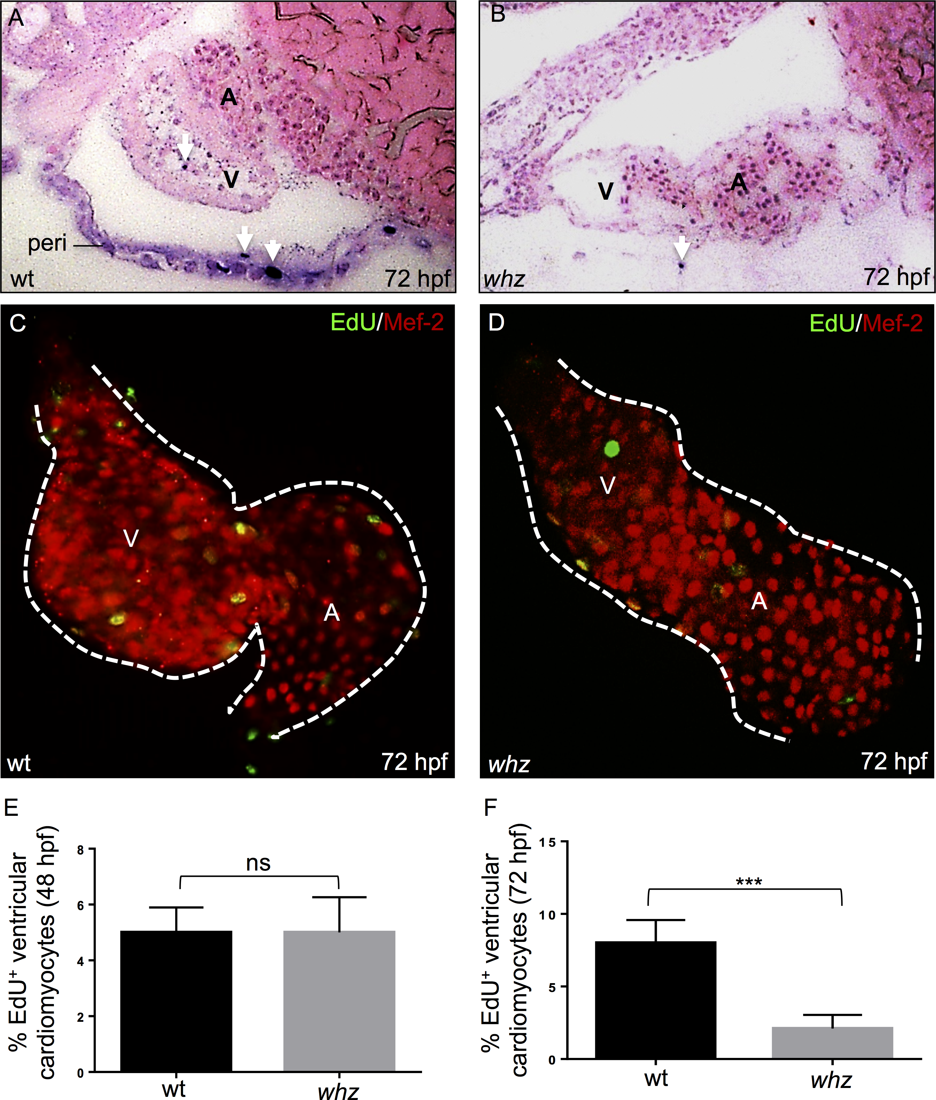
The whz mutation interferes with cardiomyocyte proliferation.
(A, B) TUNEL stainings of embryonic zebrafish hearts at 72 hpf show no difference in the number of apoptotic cardiomyocytes in wt (A) and whz (B) mutant embryos. TUNEL positive cells in the pericardium (peri) and ventricles are marked by arrows. (C-F) Dissected wt (C) and whz mutant (D) hearts at 72 hpf, stained against MEF-2 (red) after incorporation of 5-ethynyl-2'-deoxyuridine (EdU; green) to visualize cardiomyocyte proliferation. At 48 hpf, proliferation of ventricular cardiomyocytes appears unaltered between wt and whz mutant hearts (sib: 5±2% SD and whz: 4±2% SD, n = 10; p>0.05) (E), whereas cardiomyocyte proliferation in whz mutant ventricles is significantly reduced compared to wt at 72 hpf (sib: 8±2% SD, whz: 2±2% SD, n = 10, p = 0.0001) (F).