
IMAGE
Fig. 1
- ID
- ZDB-IMAGE-171201-27
- Publication
- Khatri et al., 2016 - Down-regulation of coasy, the gene associated with NBIA-VI, reduces Bmp signaling, perturbs dorso-ventral patterning and alters neuronal development in zebrafish
- All Figures
- Figures for Khatri et al., 2016
Image
|
Figure Caption
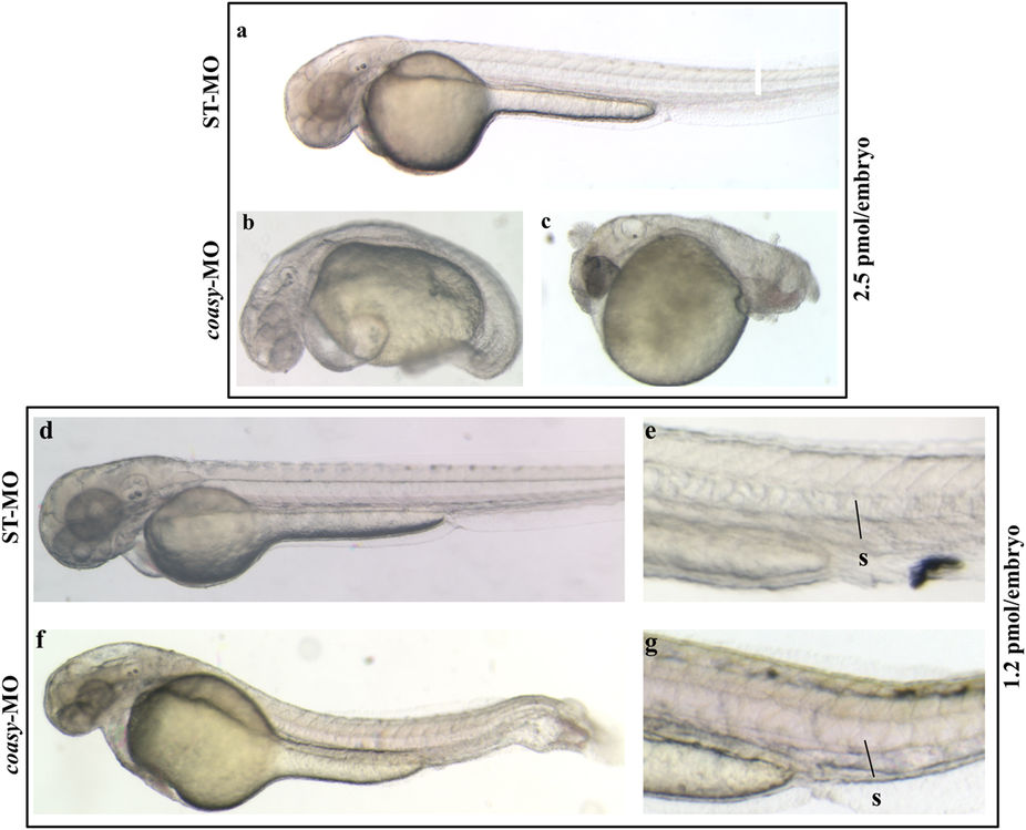
Fig. 1
Phenotypic changes due to coasy-MO microinjection.
Representative images of embryos injected with 2.5 or 1.2 pmol/embryo of ST-MO (a,d,e) or coasy-MO (b,c,f,g) at 48 hpf. Embryos injected with high doses of coasy-MO showed a severely aberrant phenotype [(B, 64%) and (C, 36%)]. Embryos injected with 1.2 pmol/embryo of coasy-MO (f,g) showed a milder phenotype.
Figure Data
Acknowledgments
This image is the copyrighted work of the attributed author or publisher, and
ZFIN has permission only to display this image to its users.
Additional permissions should be obtained from the applicable author or publisher of the image.
Full text @ Sci. Rep.