
Fig. 7
- ID
- ZDB-IMAGE-171113-6
- Publication
- Sánchez et al., 2016 - Mechanosensory organ regeneration in zebrafish depends on a population of multipotent progenitor cells kept latent by Schwann cells
- All Figures
- Figures for Sánchez et al., 2016
|
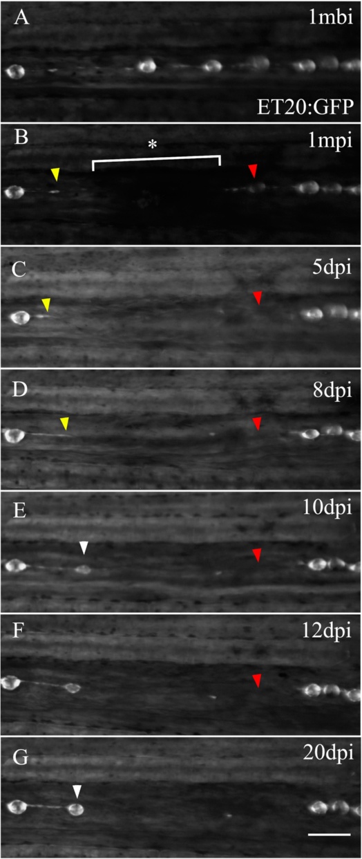
Fig. 7
Adult caudal lateral line regeneration after electroablation. A 6-month-old tg(et20:GFP) fish was electroablated in the caudal fin lateral line by applying two pulses of 2 s duration and 25 μA current intensity (n = 60, three independent experiments). Images of the injury were taken beginning from 1-minute post injury (1 mpi) to 20 days post injury (dpi). The image in a was taken at 1 mbi to show the original position of the neuromasts. The asterisk and bracket in b show the extent of the damage. c, d The yellow arrowhead points to the rostral most interneuromastic cells located immediately proximal to the damage. The red arrowheads in each panel show the disappearance of the nearest neuromast located caudal to the injury. At 10 dpi, ET20+ cells accumulated (e, white arrowhead) and at 20 dpi had matured into a neuromast (g, white arrowhead). The position where regenerated neuromasts appeared did not recapitulate the original distribution of neuromasts before injury. Scale bar a–g: 100 μm