
Image
|
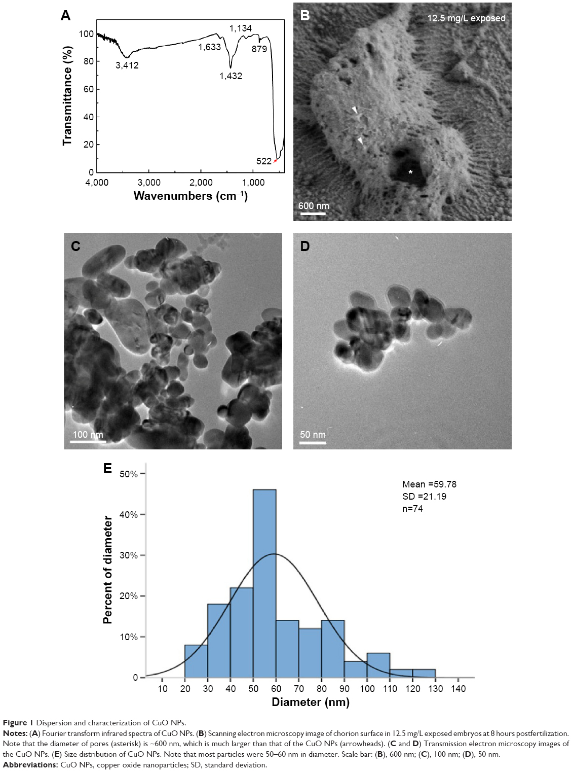
Figure Caption
Fig. 1
Dispersion and characterization of CuO NPs.
Notes: (A) Fourier transform infrared spectra of CuO NPs. (B) Scanning electron microscopy image of chorion surface in 12.5 mg/L exposed embryos at 8 hours postfertilization. Note that the diameter of pores (asterisk) is ~600 nm, which is much larger than that of the CuO NPs (arrowheads). (C and D) Transmission electron microscopy images of the CuO NPs. (E) Size distribution of CuO NPs. Note that most particles were 50–60 nm in diameter. Scale bar: (B), 600 nm; (C), 100 nm; (D), 50 nm.
Abbreviations: CuO NPs, copper oxide nanoparticles; SD, standard deviation.
Acknowledgments
This image is the copyrighted work of the attributed author or publisher, and
ZFIN has permission only to display this image to its users.
Additional permissions should be obtained from the applicable author or publisher of the image.
Full text @ Int. J. Nanomedicine