
|
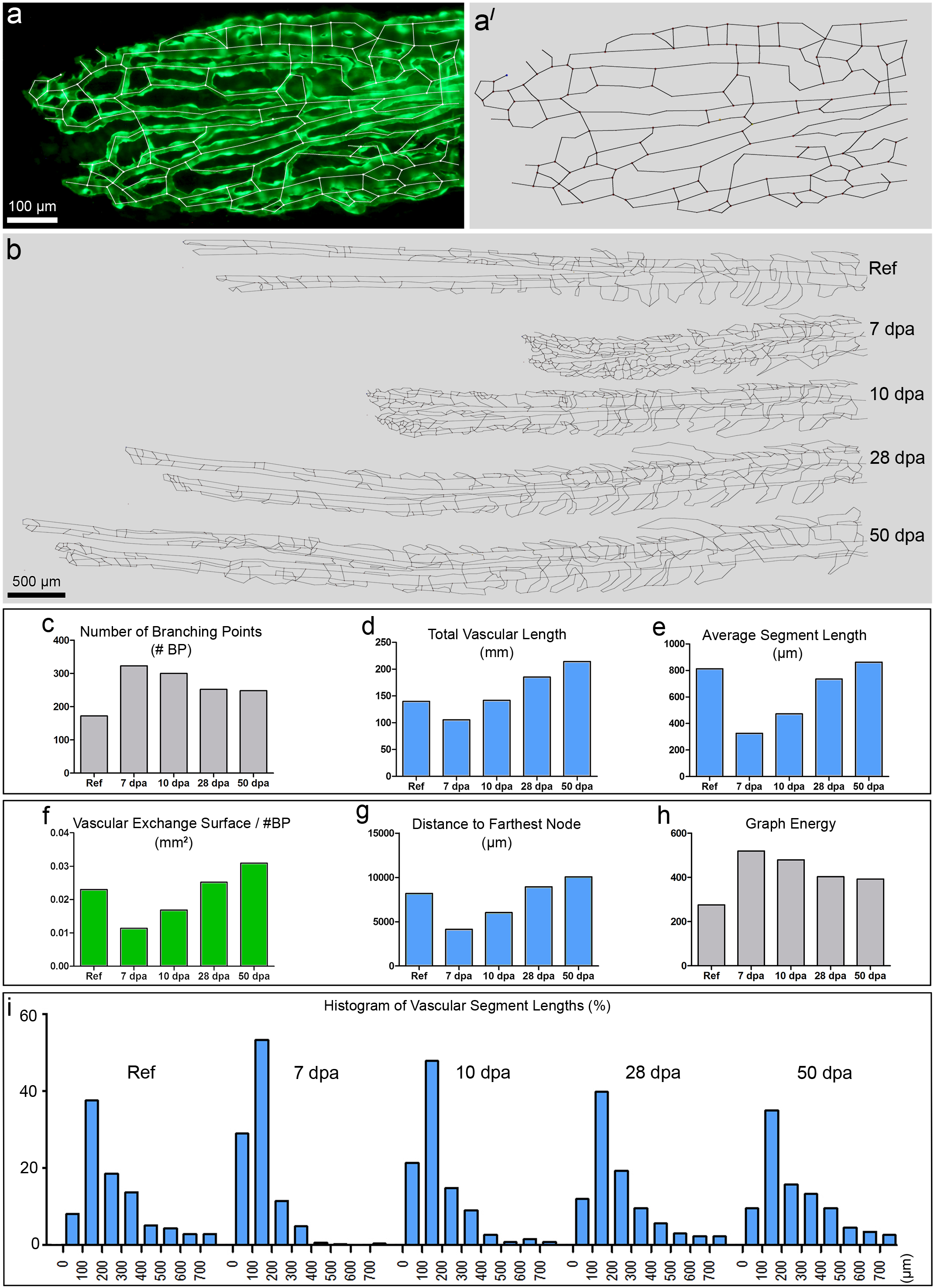
Fig. 3
Skeletonization of the vasculature of the 4th fin ray and its quantification.
a displays the distal vasculature of the 4th fin ray with overlaid skeleton. a' displays the same skeleton without the original image. b represents the skeletons of regenerated parts of the same 4th ray at different time points (reference, 7, 10, 28 and 50 dpa). The graphs in c-i display dynamics of the parameters used for quantitative analysis of the skeletons at different time points: number of branching points (c), total vascular length (d), average segment length (e), and vascular exchange surface per branching point (f), distance to farthest node (g), graph energy (h). The histograms in i showed a clear left shift at 7 dpa and 10 dpa due to the higher number of branching points and respectively shorter vascular segment lengths.