
|
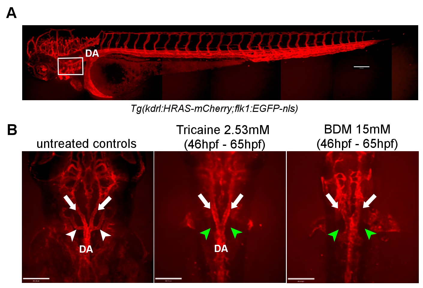
Fig. S4
Formation of the AA5x vessel in blood flow dependent.
(A) Vascular anatomy of a zebrafish embryo at 3dpf. White rectangle indicates the location of aortic arches. DA denotes dorsal aorta. Scale bar = 200μm. (B) Formation of AA5x vessels in unaffected in the WT embryos (untreated controls) as indicated by the white arrowheads. White arrows point at lateral dorsal aortae. AA5x vessel is missing in embryos treated with tricaine (middle panel) and with BDM (right figure) which both prevent blood flow (green arrowheads). Formation of lateral dorsal aortae is unaffected (white arrows). These findings confirm the previously published data [18]. Scale bar = 70μm.