
Image
|
Figure Caption
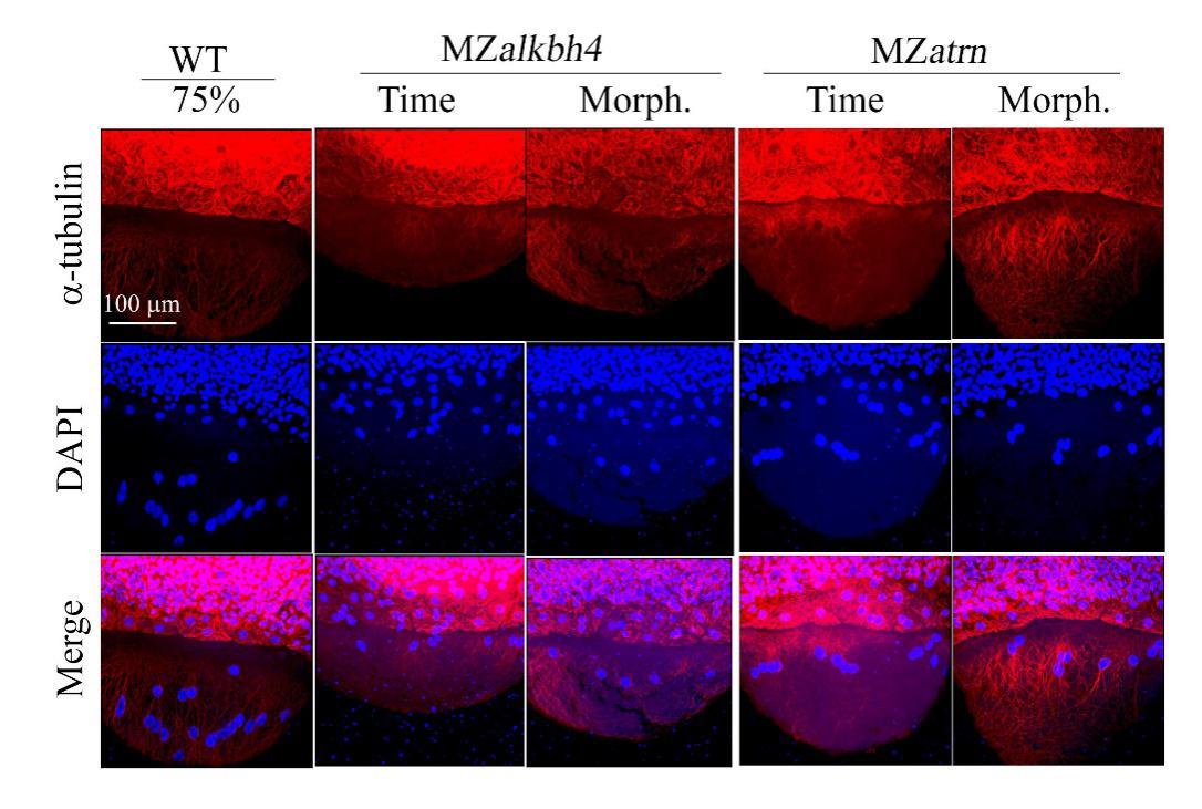
Fig. S4
Microtubule arrays in MZalkbh4 and MZatrn mutant embryos are not significantly altered. Confocal images of anti-α-tubulin and DAPI stained embryos at 75% epiboly stages. The mutant embryos were collected at both the same time point and comparable morphological stages compared with wild-type embryos. Scale bar: 100 µm.
Acknowledgments
This image is the copyrighted work of the attributed author or publisher, and
ZFIN has permission only to display this image to its users.
Additional permissions should be obtained from the applicable author or publisher of the image.
Full text @ Int. J. Biol. Sci.