
Image
|
Figure Caption
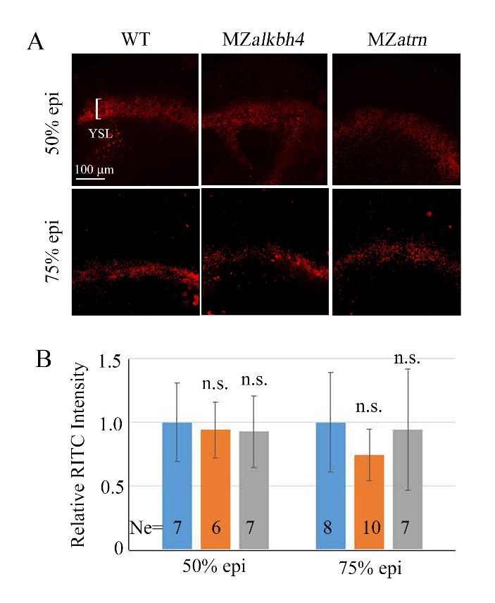
Fig. S3
E-YSL endocytosis is not affected in MZalkbh4 or MZatrn mutant embryos. (A) Fluorescence images of live embryos showing endocytic vesicles in red in the E-YSL. Embryos at 50% and 75% epiboly stages were incubated in 10 mg/ml Rhodamine B isothiocyanate-Dextran for 30 minutes, and imaged by confocal microscope. (B) The quantitative data of fluorescence intensity measured by image J software are derived from (A). Ne, the number of observed embryos. n.s. indicates no significant difference. Scale bar: 100 µm in (A).
Acknowledgments
This image is the copyrighted work of the attributed author or publisher, and
ZFIN has permission only to display this image to its users.
Additional permissions should be obtained from the applicable author or publisher of the image.
Full text @ Int. J. Biol. Sci.