
Fig. 2
- ID
- ZDB-IMAGE-171018-27
- Antibodies
- Publication
- Shim et al., 2017 - Development of zebrafish medulloblastoma-like PNET model by TALEN-mediated somatic gene inactivation
- All Figures
- Figures for Shim et al., 2017
|
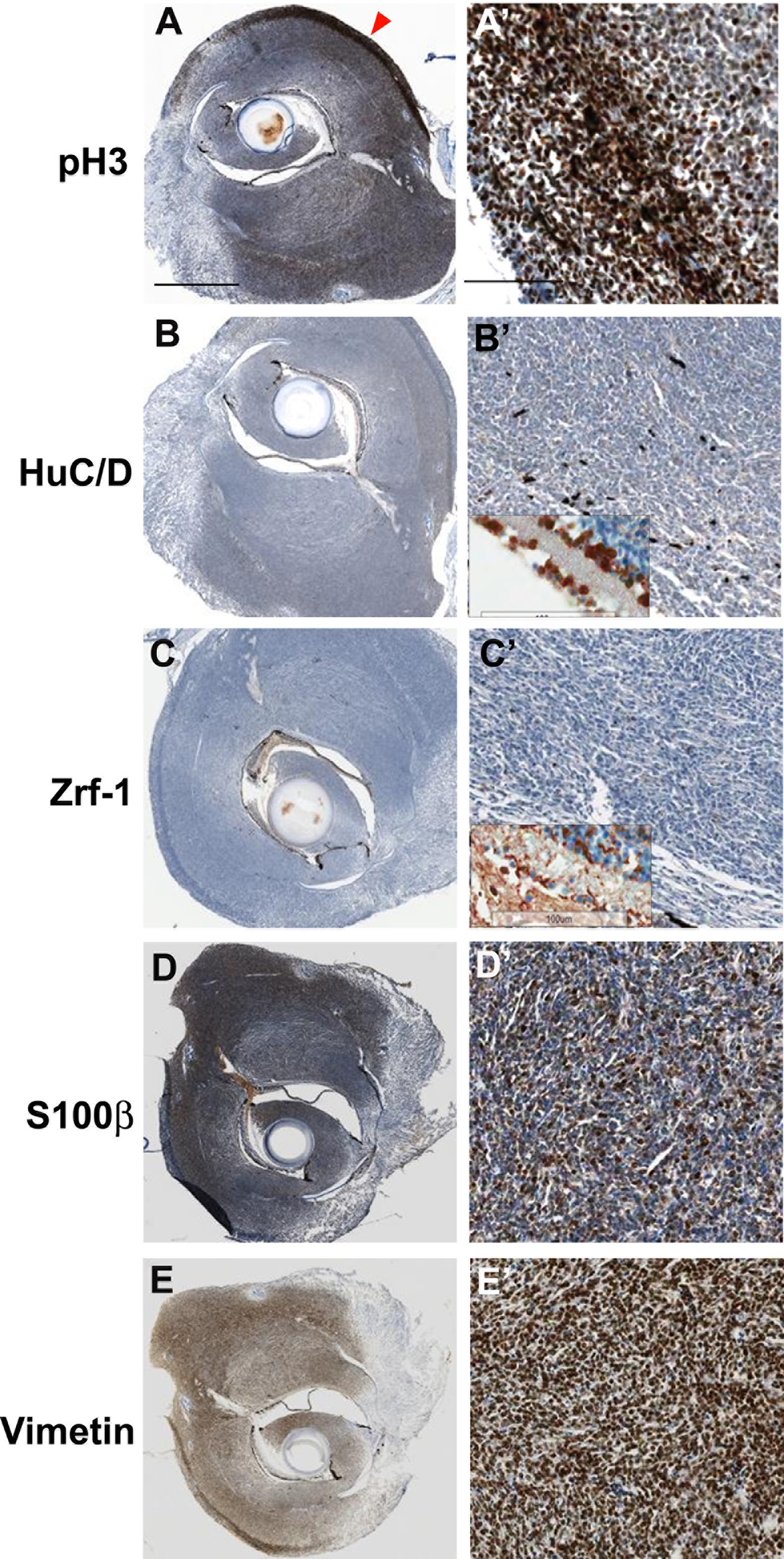
Fig. 2
Immunohistochemical analysis of tumors from cdkn2a/b TALENs mRNA injected tp53 mutant zebrafish. (A) (A') Immunostaining with anti- phospho-Histone 3 antibidy. The tumor tissue from cdkn2a/b TALENs mRNA injected tp53 mutant zebrafish is highly mitotic. The phospho-Histone 3 positive mitotic cells were more evident in the outer region of tumor tissue (red arrowhead in A). (B) (B') Neuronal signature that was visualized by immunostaining with anti-HuC/D antibody was scarcely detected in densely cellular region of tumor tissue. (C) (C') Immunostaining with zrf-1 which detect zebrafish GFAP protein. The tumor tissue induced with cdkn2a/b TALENs mRNA also did not show any glial characteristics. Normal staining of anti-HuC/D and zrf-1 antibody could be observed in retinal layers near to tumor tissues (Inset box in B' and C'). (D) (D') Epithelioid MPNST marker, S100β, is strongly expressed in tumor tissue. (E) (E') Mesenchymal marker, Vimentin, is also positively stained in tumor tissue. Scale bars: 1 mm (A) and 100 μm (A').