
Fig. 6
- ID
- ZDB-IMAGE-171016-4
- Publication
- Hübner et al., 2017 - Wnt Signaling Positively Regulates Endothelial Cell Fate Specification in the Fli1a-Positive Progenitor Population via Lef1
- All Figures
- Figures for Hübner et al., 2017
|
Fig. 6
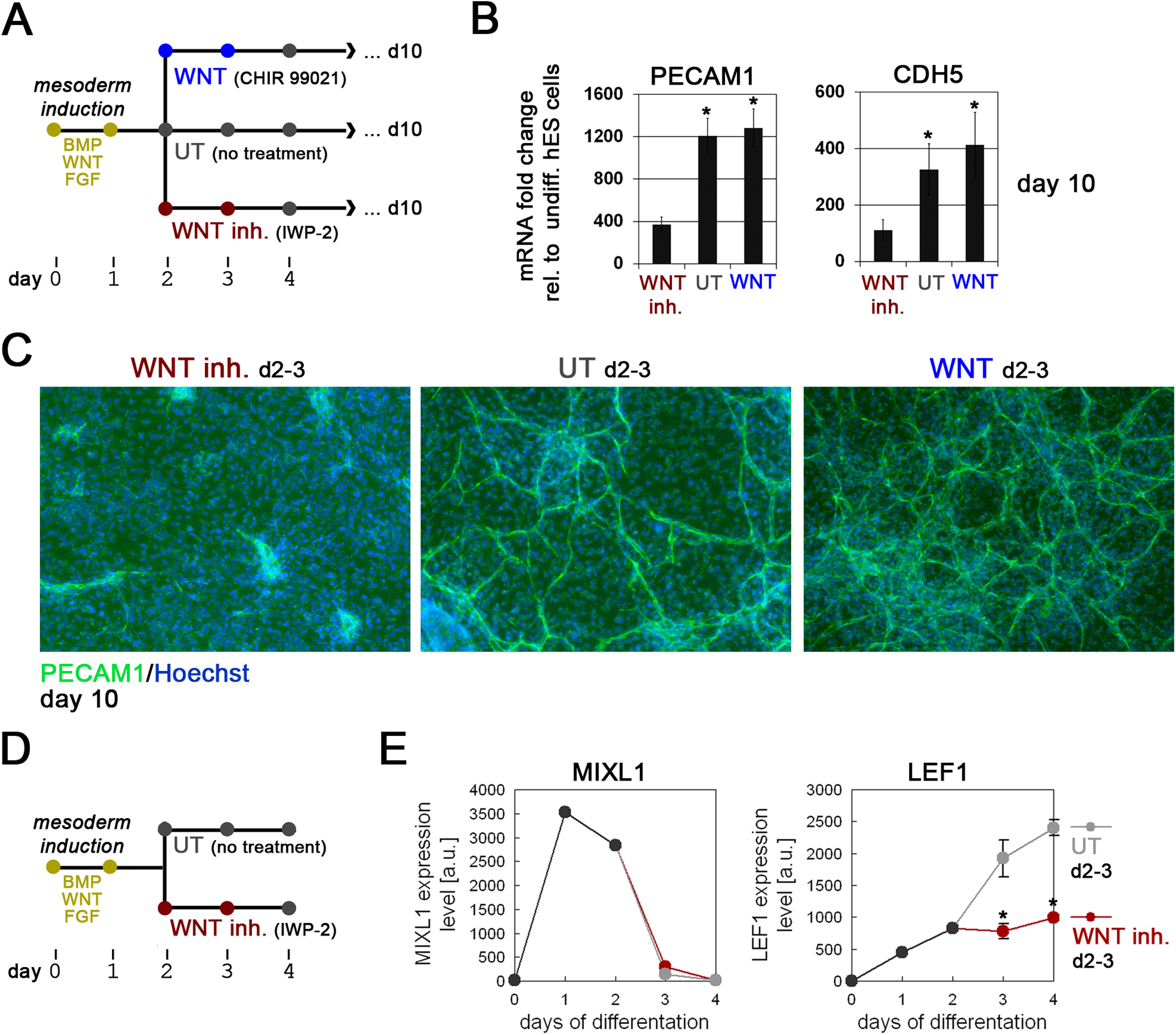
Wnt signaling promotes EC differentiation in human ES cells. (A) Differentiation protocol for investigating the effects of Wnt inhibition after an initial treatment with BMP, Wnt, and FGF to induce a mesodermal progenitor state. (B) RT-qPCR analysis of endothelial markers PECAM1 and CDH5 on day 10 of differentiation, using distinct differentiation conditions illustrated in A. (C) Immunocytochemical analysis of PECAM1 expression on day 10 using distinct differentiation conditions illustrated in A. (D-F) Gene expression time-course analysis of the primitive streak marker MIXL1 (E) as well as LEF1 (F) (microarray data). Note the decrease in LEF1 expression by inhibition of autocrine WNT signaling on days 2 and 3.
Reprinted from Developmental Biology, 430(1), Hübner, K., Grassme, K.S., Rao, J., Wenke, N.K., Zimmer, C.L., Korte, L., Mu Ller, K., Sumanas, S., Greber, B., Herzog, W., Wnt Signaling Positively Regulates Endothelial Cell Fate Specification in the Fli1a-Positive Progenitor Population via Lef1, 142-155, Copyright (2017) with permission from Elsevier. Full text @ Dev. Biol.