
|
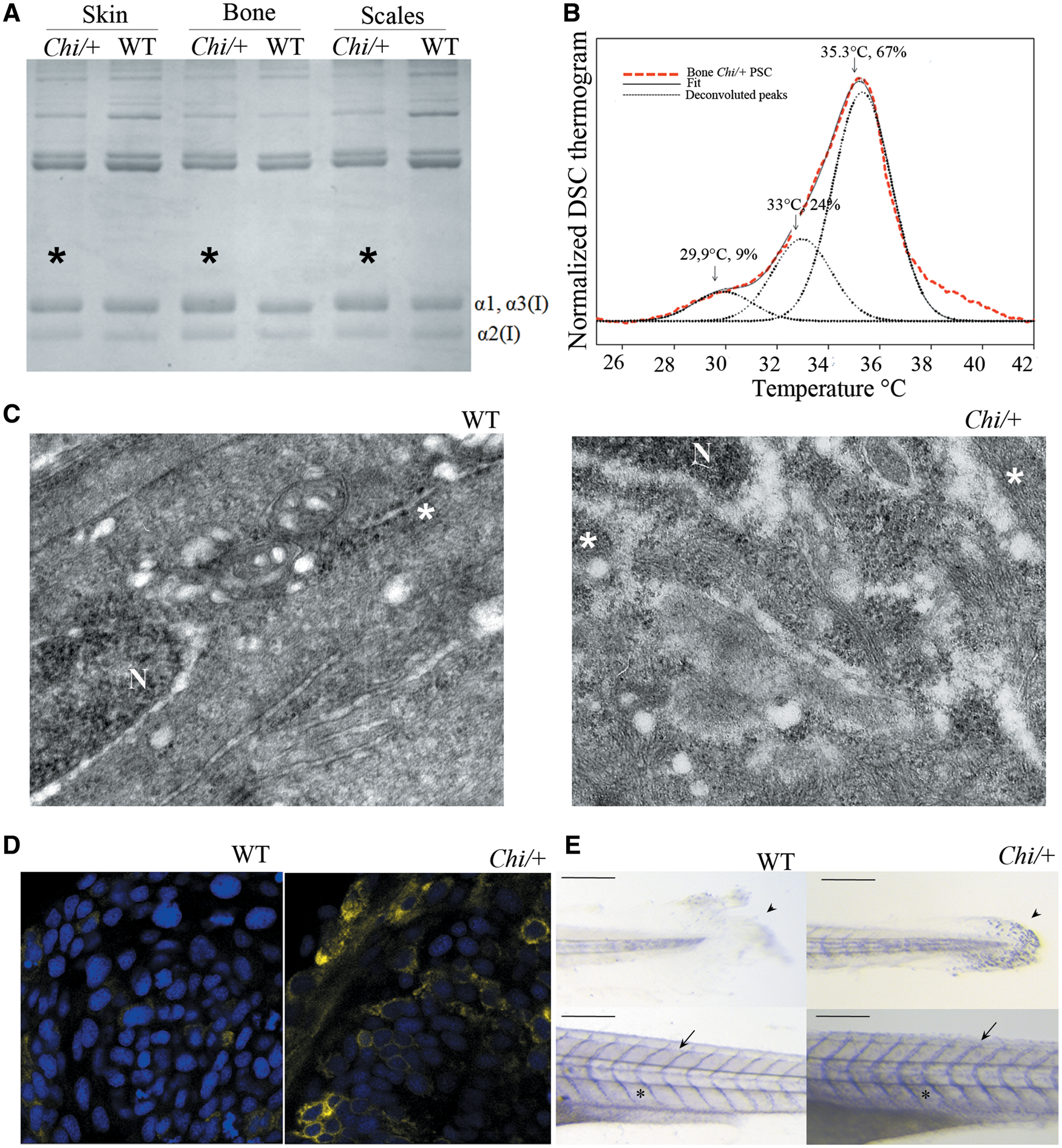
Fig. 3
Biochemical and cellular evaluation of Chi/+ collagen type I. (A) SDS-PAGE of collagen type I extracted from skin, bone and scales of WT and Chi/+ adult fish. Mutant collagen is characterized by broader α bands (asterisks), indicating collagen type I overmodification. (B) Representative DSC thermogram of collagen extracted from bone of Chi/+ adult fish. The presence of three melting temperature (Tm) peaks is evident from the deconvolution analysis of the thermogram. Similar result was obtained for collagen extracted from skin and scales (data not shown). (C) Transmission electron microscopy image of the caudal fin of WT and Chi/+ adult fish. The enlargement of endoplasmic reticulum (ER) cisternae is evident in mutant fish. N: nucleus; *: rough endoplasmic reticulum (ER). Magnification 12000X. (D) Confocal microscopy images of 24 hpf embryos WT and Chi/+ injected with mRNA expressing yellow fluorescent protein in the ER. A stronger signal in the mutant sample is demonstrated. Magnification 40X, zoom 4X. (E) Whole mount immunohistochemistry of 5 dpf WT and Chi/+ embryos using Hsp47b antibody. A stronger signal is evident in tail, skin and intersomitic space in mutant larvae. Arrow: intersomitic space, arrowhead: tail; asterisk: skin. Scale bar: 200 µm.