
|
Fig. 1
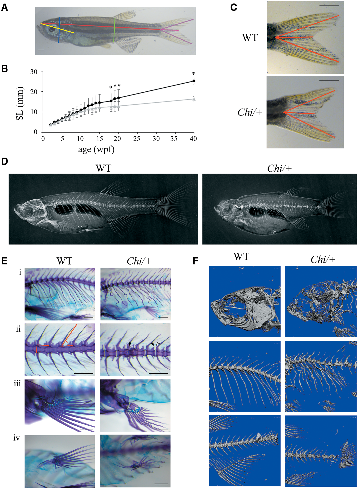
Morphological characterization of adult (Chi/+) zebrafish. (A) Morphometric parameters measured in adult fish. Standard length (SL) in red, height at anterior of anal fin (HAA) in green, height at eye (HE) in blue, snout-operculum length (SOL) in yellow, major axes of the caudal fin in purple, minor axis of the caudal fin in pink. (B) The growth curve shows that mutant fish (n = 15, grey triangles) are significantly smaller than WT (n = 12, black squares) from the 18th week post-fertilization (wpf). SL: standard length. *P < 0.05. (C) Representative image of WT and Chi/+ caudal fin. The caudal fin of mutant fish has less pronounced lobes than the WT fin. The distances measured as described in Materials and Methods are highlighted in red. Scale bar: 1 mm. (D) WT and Chi/+ X-rays show severe alterations in the skeleton of mutant fish. (E) Skeleton magnifications of 3 months old WT and Chi/+ fish stained with alcian blue and alizarin red. Major and minor axes of the vertebral body and the slope of the neural spine are indicated (ii). Deformed ribs with bone calli (i), fused vertebrae (ii, arrow), misshapen prezygapophysis and post-zygapophysis (ii, white and black arrowheads), deformed pectoral (iii) and pelvic fins (iv) are evident in mutant fish. Scale bar: 500 µm. (F) µCT of 3 months old WT and Chi/+ fish reveals the severe under mineralization of mutant animals.