
Fig. S4
- ID
- ZDB-IMAGE-170927-20
- Publication
- Madigan et al., 2017 - A Macrophage Response to Mycobacterium leprae Phenolic Glycolipid Initiates Nerve Damage in Leprosy
- All Figures
- Figures for Madigan et al., 2017
|
Fig. S4
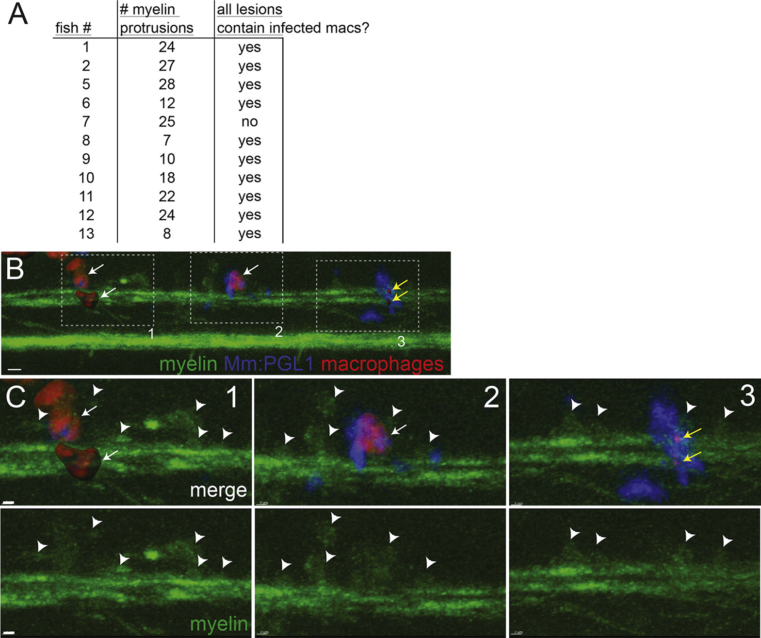
Association of Infected Macrophages with Demyelinating Lesions, Related to Figure 5I
At 2 days post-infection (5 dpf), nerve lesions were identified in M. marinum:PGL-1-infected mbp:eGFP-CAAX; mpeg1:Brainbow larvae (Figure 5I).
(A) Each lesion was scored for presence or absence of infected macrophages; association of infected macrophages with myelin protrusions was deemed significant (p = 0.01) with a two-tailed binomial test.
(B) Lesion from fish #7 (A), which had 3 demyelinating lesions (dashed boxes). For clarity, red macrophages or fragments of their red membrane are shown as rendered objects; green myelin and blue bacteria are show as the unmodified fluorescent images. 10 μm bar.
(C) Insets of each lesion from B, showing myelin protrusions and colocalized, infected macrophages that are still intact (1 and 2) or fragmented (3) and dead. Arrowheads indicate myelin protrusions; for clarity, not every myelin protrusion that was scored is indicated with an arrowhead. White arrows, intact macrophages; yellow arrows, fragments of dsRed-positive macrophage membrane. 10 μm bars.