
|
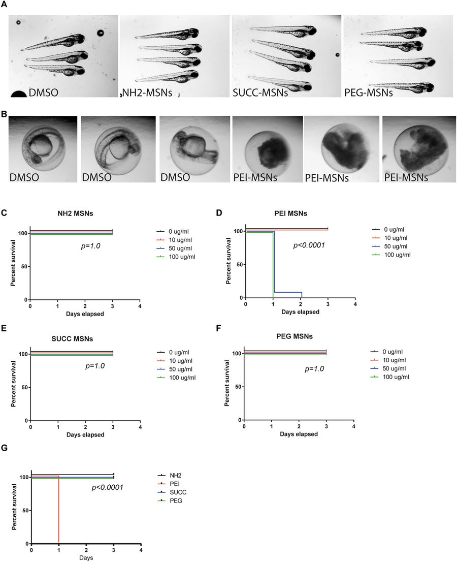
Fig. 2
Surface functionalization affects overall toxicity of MSNs. (A) Zebrafish embryos at 72 hpf treated with the DMSO vehicle, NH2-, SUCC- and PEG-MSNs at a concentration of 50 μ/ml. Treatment was initiated at 8hpf. The embryos are anesthesized and mounted in 2%methyl cellulose. (B) Zebrafish embryos at 48 hpf treated the DMSO vehicle or 50 μg/ml PEI-MSNs. Treatment was initiated at 8hpf. (C–F) Percentage of survival of embryos at 24hpf, 48 hpf and 72hpf upon treatment with (C) NH2-, (D) PEI-, (E) SUCC- and (F) PEG-MSNs at denoted concentrations. The treatment was initiated at 8hpf. N = 24 embryos/treatment, except in PEI-MSN 10 μg/ml (n = 25) and SUCC-MSN 100 μg/ml (n = 25). Each embryo was followed throughout the experiment and score live or dead on each day (G) Comparison of survival between nanoparticles at 100 μg/ml concentration.