
|
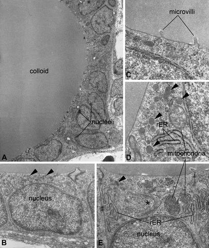
Fig. 1
Ultrastructure of thyroidal tissue in control zebrafish (Danio rerio). The epithelium encloses an evenly stained colloid devoid of inclusions (A). The nucleus is basally located and most organelles can be found in apical position (B, E). Mitochondria appear spherically to ovally shaped; the rough endoplasmic reticulum and Golgi fields (*) are of cistern-like appearance (D, E). At the apical pole of thyrocytes, few electron-dense lysosomes are detectable (▸), and at the border to the colloid, some microvilli are detectable (B, C, D, E). Magnifications: A: 2,000×; B: 10,000×; C: 12,500×; D: 40,000×; E: 31,500×.