
Fig. 3
- ID
- ZDB-IMAGE-170921-55
- Publication
- Sedykh et al., 2017 - Zebrafish zic2 controls formation of periocular neural crest and choroid fissure morphogenesis
- All Figures
- Figures for Sedykh et al., 2017
|
Fig. 3
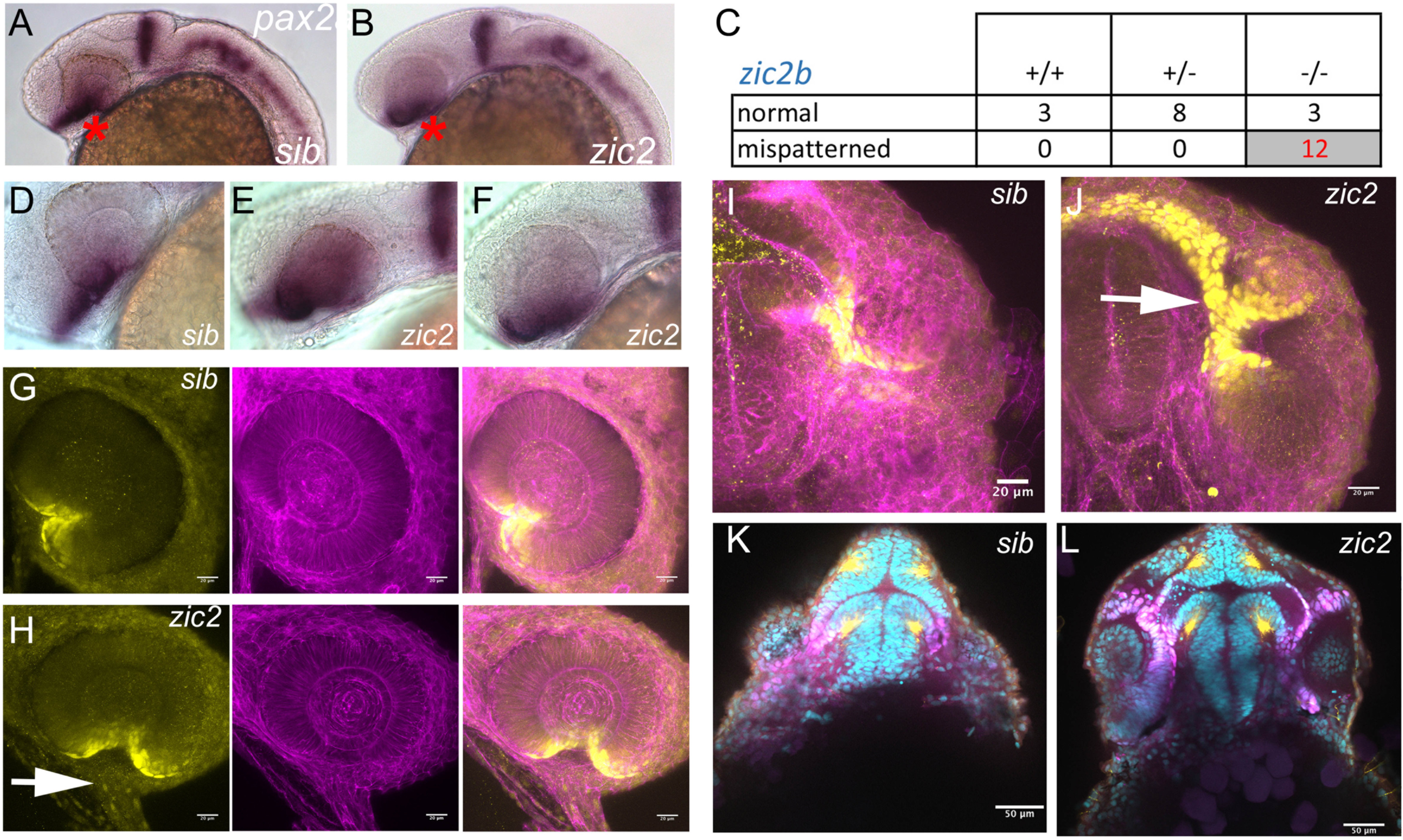
Pax2a expression is aberrant in MZ-zic2 mutants at 1 dpf. pax2a expression at 1 dpf was visualized in embryos derived from zic2agbt133/+; zic2buw1116/+ parents using WISH (A-F) or in progeny of zic2agbt133/+; zic2buwt104 parents using immunohistochemistry (G-L). A: normal pax2a expression in the ventral retina (*). B: mispatterned pax2a expression (*) was observed in 12 out of 103 embryos (12%, 2 expts.). C: Only zic2b homozygous embryos exhibit pax2a mispatterning. zic2a genotype was not tested because PCR genotyping was not robust after WISH. D-F: Embryos with mispatterned pax2a expression also exhibit coloboma, indicative of homozygosity for zic2agbt133. G, H: confocal stacks through representative retina of normal (G) and zic2 mutant (H) retina. I, J: confocal stacks through the ventral aspects of a normal (I) and zic2 mutant (J) diencephalon and retina. Arrowheads in H, J point to the aberrant optic stalk. In G-J, yellow = Pax2a, magenta = F-actin cytoskeleton visualized by phalloidin. K, L: single confocal sections through representative normal (K) and zic2 mutant (L) embryos, imaged ventrally at the level of choroid fissure. magenta = Pax2a; yellow = acetylated tubulin; cyan = nuclei visualized by DAPI. Embryos are shown in lateral views, anterior to the left (A-F) or anterior to the right (G, H); in ventral views with anterior at the top (I-L).
Reprinted from Developmental Biology, 429(1), Sedykh, I., Yoon, B., Roberson, L., Moskvin, O., Dewey, C.N., Grinblat, Y., Zebrafish zic2 controls formation of periocular neural crest and choroid fissure morphogenesis, 92-104, Copyright (2017) with permission from Elsevier. Full text @ Dev. Biol.