
IMAGE
Fig. S11
- ID
- ZDB-IMAGE-170908-18
- Publication
- Breau et al., 2017 - Extrinsic mechanical forces mediate retrograde axon extension in a developing neuronal circuit
- All Figures
- Figures for Breau et al., 2017
Image
|
Figure Caption
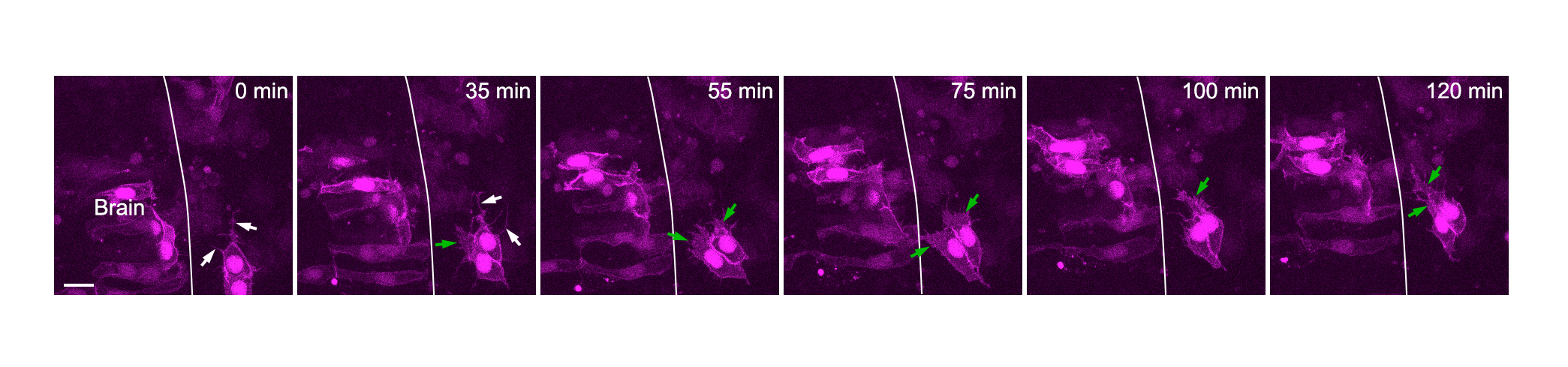
Fig. S11
Pacodal cells exhibit filopodia and lamellipodia-like protrusions during convergence. Live imaging on a wild type embryo transplanted with mbCherry and H2B-RFP co-expressing cells, from 12s onwards. Note the filopodia in the front of posterior placodal cells converging towards the center (white arrows), but also the presence of large lamellipodia-like protrusions (green arrows). The white line indicates the brain surface. XY dorsal view, anterior to the top, scale bar: 5 µm.
Acknowledgments
This image is the copyrighted work of the attributed author or publisher, and
ZFIN has permission only to display this image to its users.
Additional permissions should be obtained from the applicable author or publisher of the image.
Full text @ Nat. Commun.