
Fig. 5 S2
- ID
- ZDB-IMAGE-170907-8
- Publication
- Sugimoto et al., 2017 - Dissection of zebrafish shha function using site-specific targeting with a Cre-dependent genetic switch.
- All Figures
- Figures for Sugimoto et al., 2017
|
Fig. 5 S2
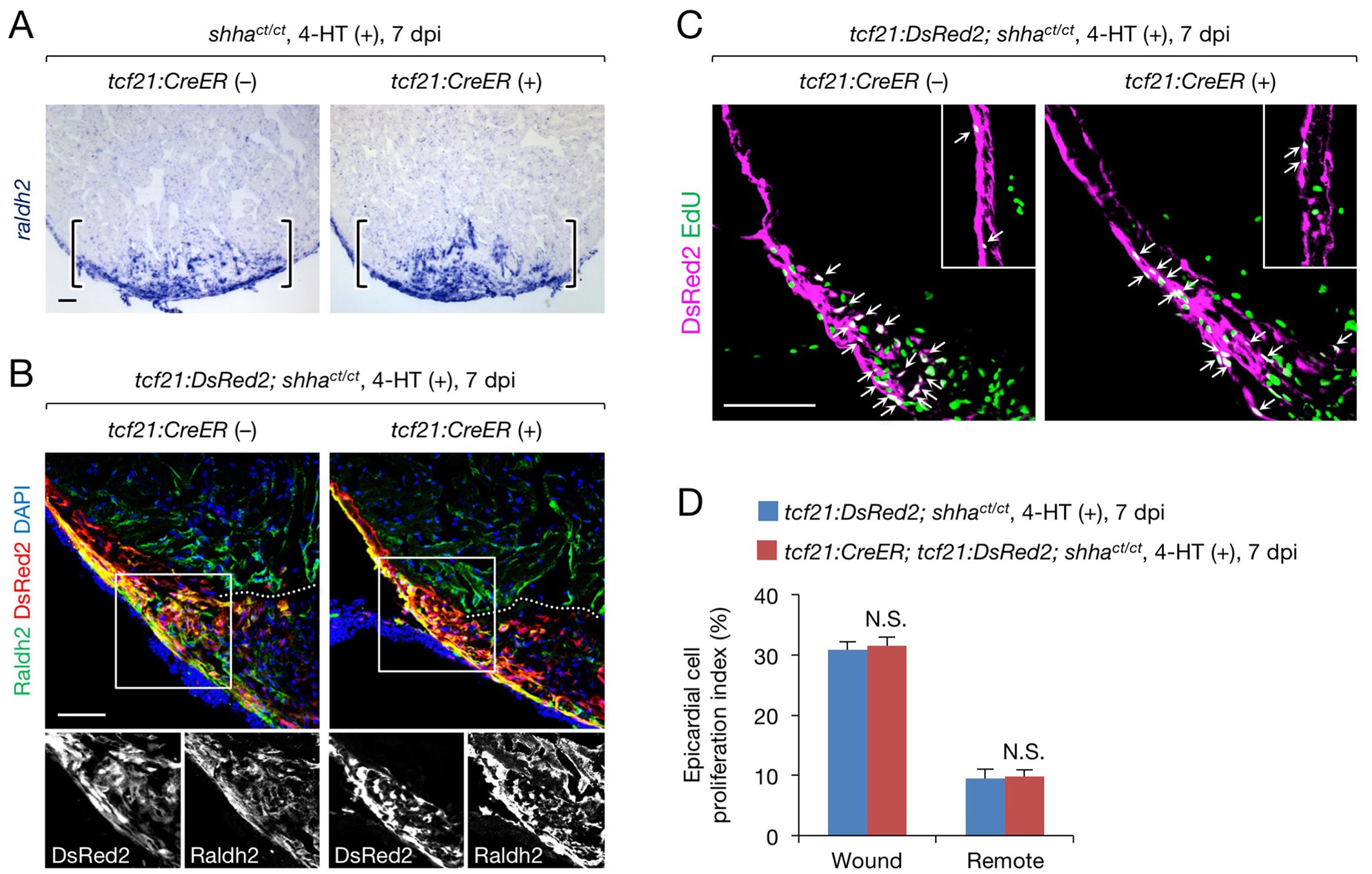
A redundant role for epicardial shha in epicardial migration and proliferation during heart regeneration.
(A) In situ hybridization analysis of raldh2 expression in sections obtained from 4-HT-treated 7 dpi shhact/ct (control) and tcf21:CreER; shhact/ct hearts. Brackets, injury site. (B) Immunofluorescence staining of Raldh2 and DsRed2 using sections obtained from 4-HT-treated 7 dpi tcf21:DsRed; shhact/ct (control) and tcf21:DsRed; tcf21:CreER; shhact/ct hearts (right). Single-channel images of the rectangle are shown at the bottom. Dotted line, approximate amputation plane. (C) Immunofluorescence staining of DsRed2 and EdU in sections obtained from 4-HT-treated 7 dpi tcf21:DsRed; shhact/ct (control) and tcf21:CreER; tcf21:DsRed; shhact/ct hearts. Inset, non-injured area. Arrows indicate proliferating tcf21+ epicardial cells, which were defined as epicardial cells colabeled DsRed2 and EdU. (D) Quantification of epicardial cell proliferation in the sections obtained from 4-HT-treated 7 dpi tcf21:DsRed; shhact/ct (control) and tcf21:CreER; tcf21:DsRed; shhact/ct hearts shown in C (n = 5 each). The data represent the mean ± SEM (wound, p=0.754; remote, p=0.602; Mann–Whitney U test). N.S., not significant. Single confocal slice images are shown in B and C. Scale bar, 50 μm.