
|
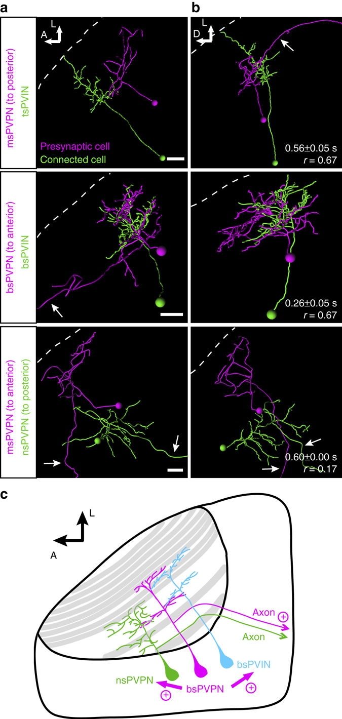
Fig. 7
Optobow reveals novel excitatory connections of projection neurons within the optic tectum. a, b Morphological reconstructions of connected tectal cells, identified using Optobow-nPA, in dorsal view (a) and transverse view (b). Cell types are indicated on the left. White dashed lines mark the skin. Arrows point to projection axons leaving the neuropil. Values for response onset time (s) and response reliability (r) are shown in b. Scale bar, 15 µm. c Model for tectal connectivity of PVPNs. In addition to an axon leaving the tectum, PVPNs make functional, excitatory connections both with PVINs and other PVPNs. bs, bistratified; ms, monostratified; ns, non-stratified; PVIN, periventricular interneuron; PVPN, periventricular projection neuron; ts, tristratified