
Fig. S10
- ID
- ZDB-IMAGE-170828-7
- Publication
- Wehner et al., 2017 - Wnt signaling controls pro-regenerative Collagen XII in functional spinal cord regeneration in zebrafish
- All Figures
- Figures for Wehner et al., 2017
|
Fig. S10
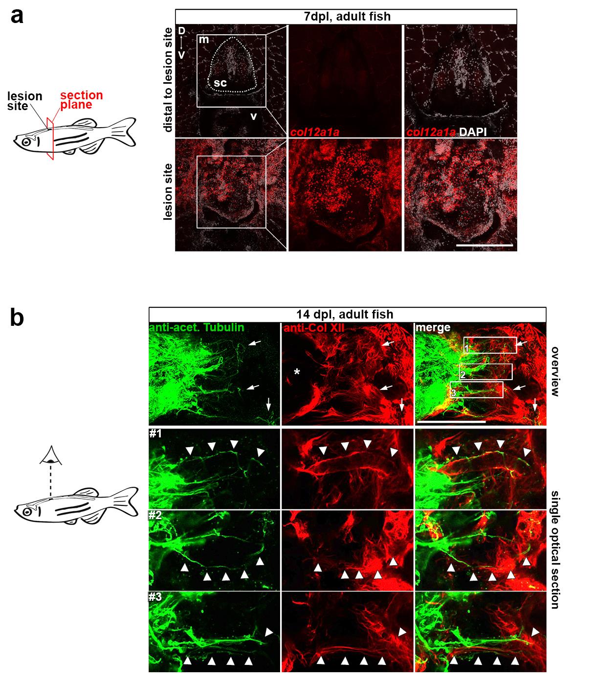
Col XII matrix deposition in a spinal lesion site is conserved across developmental stages in zebrafish.
(a) In adult zebrafish, col12a1a is strongly expressed in a spinal lesion site (bottom panel). Transcripts are undetectable in the intact spinal cord distal to the lesion site (top panel). Abbreviations: sc, spinal cord; m, muscle; v, vertebra.
(b) In adult zebrafish, regenerating axons (anti-acetylated Tubulin+) navigate a Col XII-rich lesion environment (arrows). Note that Col XII immunoreactivity is undetectable in the intact portion of the spinal cord (asterisk). Shown is a maximum intensity projection (82 μm) and single optical sections with higher magnification showing close association between axons and Col XII. Note that the trajectory of many axonal fascicles appear to follow longitudinal fibres of Col XII immunoreactive ECM material in the lesion site (arrowheads).
(a-b) Views are transversal (a; dorsal is up) or dorsal (b; rostral is left). Scale bars: 250 μm (a), 200 μm (b).