
Fig. S9
- ID
- ZDB-IMAGE-170828-6
- Publication
- Wehner et al., 2017 - Wnt signaling controls pro-regenerative Collagen XII in functional spinal cord regeneration in zebrafish
- All Figures
- Figures for Wehner et al., 2017
|
Fig. S9
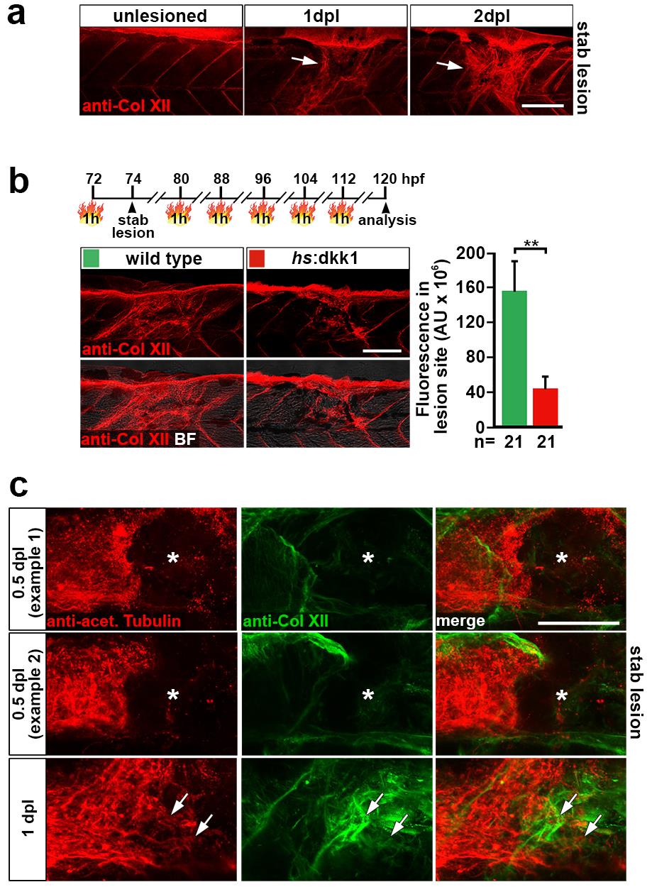
Wnt/β-catenin signaling controls Col XII matrix deposition in a spinal lesion site through which regenerating axons grow.
(a) Anti-Col XII immunoreactivity is increased in the lesion site (arrows) at 1 dpl and 2 dpl compared to adjacent unlesioned tissue and to unlesioned animals at the same trunk position.
(b) Inhibition of Wnt/β-catenin signaling via heat shock-induced systemic overexpression of the pathway antagonist dkk1 interferes with Col XII deposition in the lesion site, as determined by quantification of lesion site immunoreactivity (t-test: **P<0.01).
(c) Double immunolabelling of axons (anti-acetylated Tubulin+) and Col XII shows little to no Col XII deposition in the lesion site at 0.5 dpl (12 hpl) and axon are yet to enter the lesion site (asterisk). At 1 dpl, anti-Col XII immunoreactivity is markedly increased in the lesion site and axon have entered the Col XII-rich lesion site (arrow). Confocal depth was limited to spinal cord. Note, that the 1 dpl dataset is the same as presented in Fig. 4f.
(a-c) Views are lateral (dorsal is up, rostral is left). BF: brightfield. Scale bars: whole mounts, 100 μm (a-b) and 50 μm (c). Error bars indicate s.e.m.