
IMAGE
Fig. S14
- ID
- ZDB-IMAGE-170828-10
- Publication
- Wehner et al., 2017 - Wnt signaling controls pro-regenerative Collagen XII in functional spinal cord regeneration in zebrafish
- All Figures
- Figures for Wehner et al., 2017
Image
|
Figure Caption
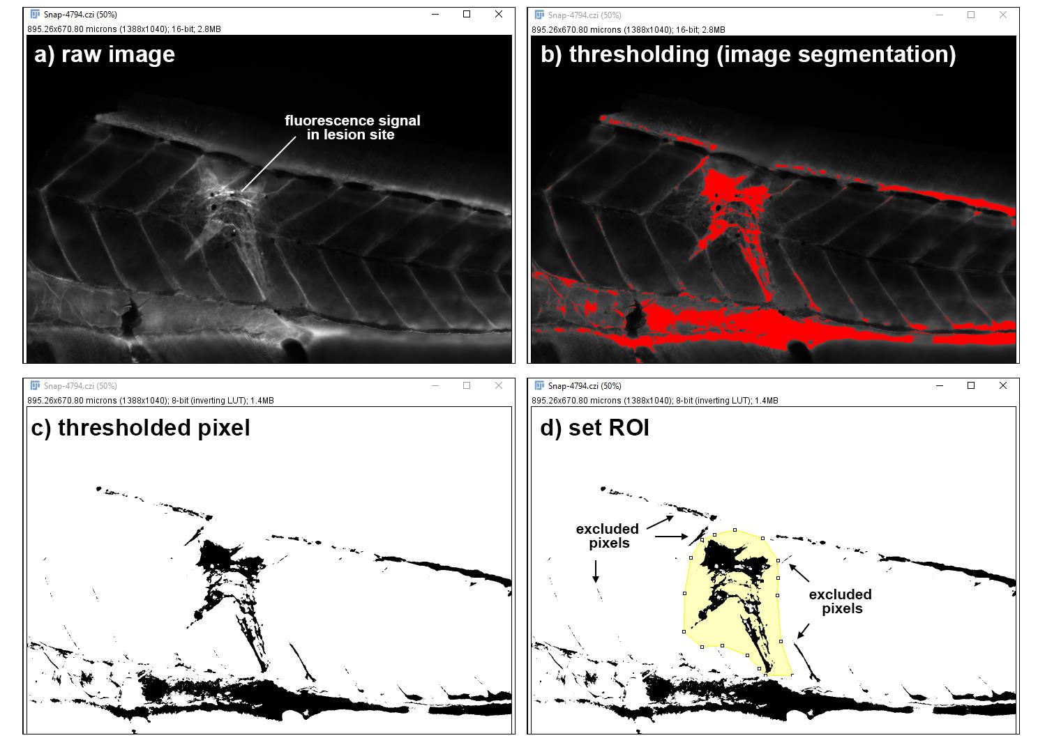
Fig. S14
Workflow for the quantitative analysis of immunohistochemistry or fluorescence in situ hybridization signals in a lesion site.
A minimum intensity threshold is applied to the raw image (a-b), limiting pixels to only those of equal or higher intensity (c). After thresholding, a ROI is defined manually in which the thresholded pixel area is measured (d). The analysis is done without knowledge of the experimental condition.
Acknowledgments
This image is the copyrighted work of the attributed author or publisher, and
ZFIN has permission only to display this image to its users.
Additional permissions should be obtained from the applicable author or publisher of the image.
Full text @ Nat. Commun.