
Fig. S2
- ID
- ZDB-IMAGE-170825-39
- Publication
- Wehner et al., 2017 - Wnt signaling controls pro-regenerative Collagen XII in functional spinal cord regeneration in zebrafish
- All Figures
- Figures for Wehner et al., 2017
|
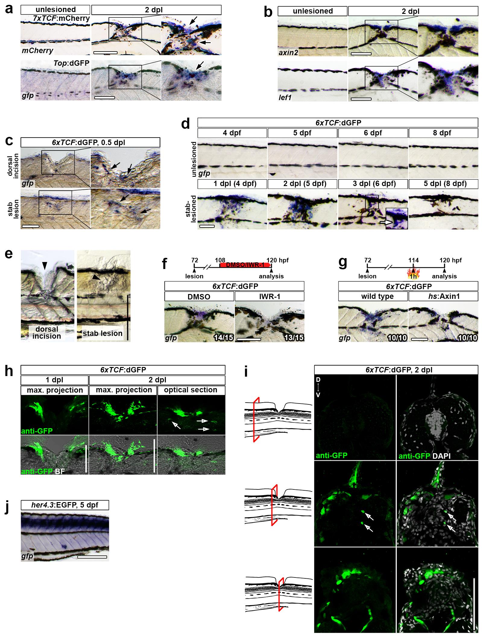
Fig. S2
Wnt/β-catenin pathway is mainly active in the spinal lesion site.
(a) Expression of the transgenic Wnt/β-catenin pathway reporter 7xTCF:mCherry and Top:dGFP is upregulated after lesion.
(b) Expression of direct Wnt target genes axin2 and lef1 is upregulated after lesion.
(c) 6xTCF:dGFP Wnt reporter activity in the spinal lesion site is detectable 12 hours (0.5 dpl) after a stab lesion or dorsal incision lesion (arrows). Note that larvae were treated with PTU to suppress pigmentation.
(d) Detection of gfp mRNA in the 6xTCF:dGFP transgenic reporter line shows transient activity of the Wnt/β-catenin pathway in the lesion site during regeneration, analysed in stab-lesioned animals.
(e) Representative images of a dorsal incision lesion or less invasive stab lesion taken immediately after lesion. Note that stab lesions do not compromise the dorsal edge of the animal, resulting in reduced injury size as compared to dorsal incision lesions. Arrowhead points to the lesion site.
(f) 6xTCF:dGFP Wnt reporter activity in the lesion site is strongly reduced upon IWR-1 treatment for 12 hours, indicating specificity of the transgenic Wnt reporter.
(g) 6xTCF:dGFP Wnt reporter activity in the lesion site is strongly reduced upon heat shock-induced axin1 overexpression for 6 hours in 6xTCF:dGFP;hs:Axin1 double transgenic animals, indicating specificity of the transgenic Wnt reporter.
(h) GFP protein is largely confined to the lesion site in 6xTCF:dGFP transgenic animals at 1 dpl and 2 dpl. A few additional cells are labelled within the presumptive spinal cord region in the periphery of the lesion site (empty arrows). Maximum intensity projections and a single optical section through the center of a whole mount larvae are shown.
(i) Anti-GFP immunohistochemistry on sections of 6xTCF:dGFP transgenic animals confirms labelling in whole mount preparations. 6xTCF:dGFP Wnt reporter activity at 2 dpl is largely confined to the lesion site. In the peripheral lesion area limited Wnt reporter activity is also detected in the spinal cord (empty arrows).
(j) Detection of gfp mRNA by in situ hybridization in 5 day-old her4.3:EGFP transgenic zebrafish, labelling ependymoradial glia cells, reveals efficient probe penetration in whole mount preparations.
(a-j) Views are lateral (a-h, j; dorsal is up, rostral is left) or transversal (i; dorsal is up). BF: brightfield. Scale bars: whole mounts, 200 μm (j,e) and 100 μm (a-d, f-h); sections, 100 μm.