
IMAGE
Fig. S14
- ID
- ZDB-IMAGE-170823-18
- Publication
- Wang et al., 2016 - Wars2 is a determinant of angiogenesis
- All Figures
- Figures for Wang et al., 2016
Image
|
Figure Caption
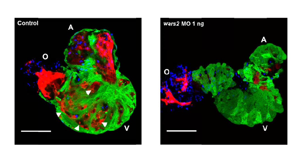
Fig. S14
Confocal images optically sectioned at a depth of 2µm into the wall of isolated hearts of 4dpf Tg(myl7:GFP;flk:dsRed) zebrafish embryos.
In control embryos there are numerous infiltrations of endothelial cells (red) interspersed between myocardial (green) and trabeculae (arrowheads), which are almost completely absent in hearts lacking Wars2. Blue: Hoechst staining. A: atrium, O: outflow tract, V: ventricle. Scale bar=50µm.
Figure Data
Acknowledgments
This image is the copyrighted work of the attributed author or publisher, and
ZFIN has permission only to display this image to its users.
Additional permissions should be obtained from the applicable author or publisher of the image.
Full text @ Nat. Commun.