
Fig. 8
- ID
- ZDB-IMAGE-170822-8
- Genes
- Publication
- Ke et al., 2017 - Gβ1 is required for neutrophil migration in zebrafish
- All Figures
- Figures for Ke et al., 2017
|
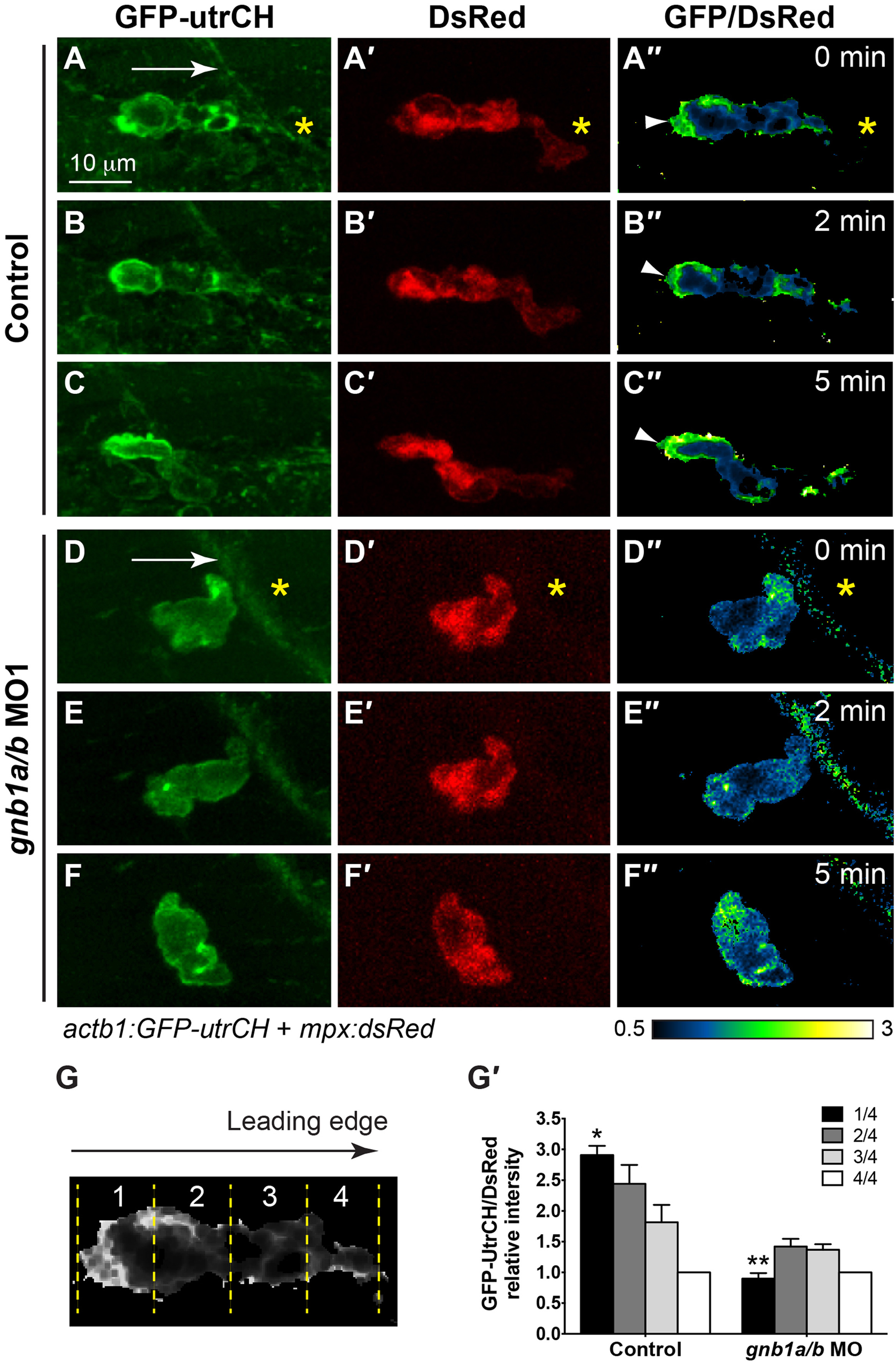
Fig. 8
Gβ1 regulates the polarity of stable F-actin accumulation in neutrophils. (A-F) Snapshots from confocal time-lapse imaging (Supplementary Movie 8) of migrating neutrophils expressing GFP-utrCH in control and gnb1a/b MO1-injected embryos following wounding. (A-F) GFP-utrCH. (A′-F′) DsRed. (G′′-L′′) Ratiometric images of GFP-utrCH/DsRed. Yellow asterisks: side nearest wound; white arrowheads: GFP-utrCH enrichment in neutrophil trailing region; white arrows, direction of migration. (G-G′) Quantification of GFP-utrCH/DsRed. (G) Illustration of the method used to quantify ratio of intensities of PHAKT-EGFP/DsRed, as described in Fig. 6G. (G′) Graph showing the ratio of intensities of GFP-utrCH/DsRed in each quarter of the cell relative to that in the fourth quarter (the leading region), in control (5 neutrophils in 5 embryos) and gnb1a/b MO1-injected (9 neutrophils in 4 embryos) embryos. * p = 0.0001 vs 1/4 in control, ** p < 0.0001 vs 1/4 in control, by t-test, two-way ANOVA; p < 0.0001 among groups in control embryos, by one-way ANOVA; p = 0.0003, among groups in gnb1a/b MO1-injected embryos, by one-way ANOVA.
Reprinted from Developmental Biology, 428(1), Ke, W., Ye, D., Mersch, K., Xu, H., Chen, S., Lin, F., Gβ1 is required for neutrophil migration in zebrafish, 135-147, Copyright (2017) with permission from Elsevier. Full text @ Dev. Biol.