Fig. S1
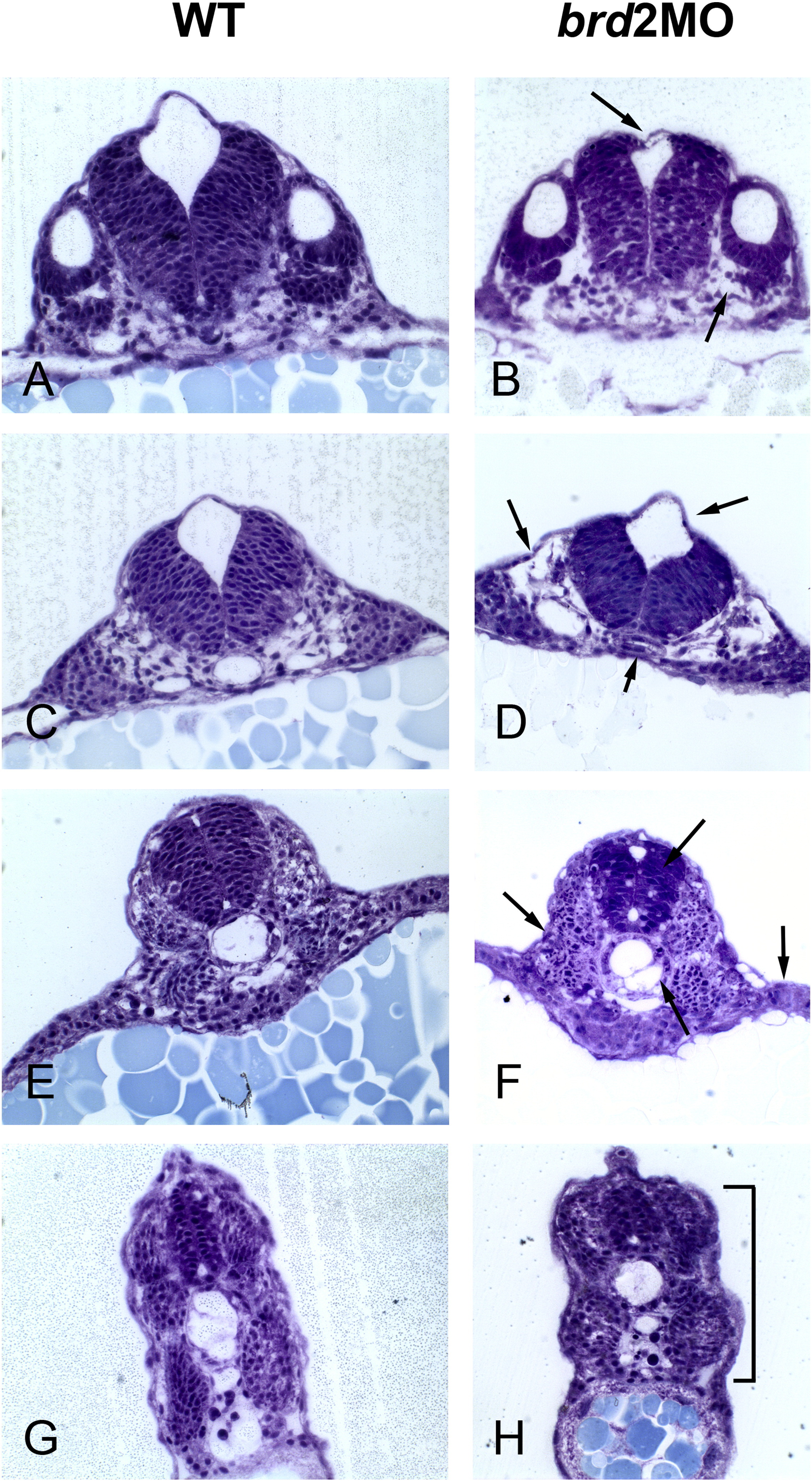
brd2aMO morphants exhibit abnormal tissue architecture in hindbrain, neural tube, somites and paraxial mesoderm. Transverse plastic sections through hindbrain (A–F) and trunk (G,H) of uninjected wildtype (WT) and 8 ng brd2aMO1-injected morphants (brd2aMO) were stained with toluidine blue and compared. A,B) show hindbrain at level of otic vesicle; C,D) and E,F) show progressively more caudal sections of hindbrain. brd2aMO1 morphants have collapsed ventricles (top arrows in B, D), disorganized, loosely packed mesoderm (bottom right arrow in B, top left arrow in D, left and right arrows in F), abnormal notochords (bottom arrows in D,F), and irregular, contracted somites (bracket in H). Although control-injected embryos were not compared directly in this analysis, the hindbrain and spinal cord irregularities we see here are corroborated by images of embryos obtained with BrdU and TUNEL staining, where samples of control-injected MOc embryos show tissue architecture similar to uninjected WT (see Results Section 2.3.2).
Reprinted from Mechanisms of Development, 146, Murphy, T., Melville, H., Fradkin, E., Bistany, G., Branigan, G., Olsen, K., Comstock, C.R., Hanby, H., Garbade, E., DiBenedetto, A.J., Knockdown of epigenetic transcriptional co-regulator Brd2a disrupts apoptosis and proper formation of hindbrain and midbrain-hindbrain boundary (MHB) region in zebrafish, 10-30, Copyright (2017) with permission from Elsevier. Full text @ Mech. Dev.

