
Fig. 5
- ID
- ZDB-IMAGE-170807-15
- Publication
- Xu et al., 2017 - Neurons secrete miR-132-containing exosomes to regulate brain vascular integrity
- All Figures
- Figures for Xu et al., 2017
|
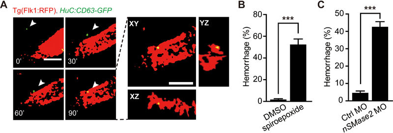
Fig. 5
Neuronal exosomes are translocated to brain ECs in larval zebrafish. (A) In vivo time-lapse confocal images showing that a GFP-positive neuronal exosome (white arrowheads) approaches and enters RFP-positive ECs in the brain. The images were taken from a 3-dpf Tg(Flk1:RFP) zebrafish larva, in which HuC:CD63-GFP was transiently expressed to label neuronal exosomes. To trace the moving GFP-positive exosome at each time point, single-slice images at different optical sections are shown at the Left. To confirm the exosome internalization into ECs, three-dimensional views of the single-slice image at 90′ are shown at the Right. (B, C) Intracranial hemorrhage effects of nSMase2 blockade with spiroepoxide (B) or nSMase2 knockdown with MO (C) in zebrafish. The nSMase2 is required for the budding of exosomes into multi-vesicular bodies. The experiments were repeated seven times in B and six times in C. Scale bar, 5 μm (A). Error bars, SEM. ***P < 0.001 (unpaired two-tailed Student's t-test for B and C).