
Fig. 7
- ID
- ZDB-IMAGE-170706-21
- Publication
- Pei et al., 2016 - Additive reductions in zebrafish PRPS1 activity result in a spectrum of deficiencies modeling several human PRPS1-associated diseases
- All Figures
- Figures for Pei et al., 2016
|
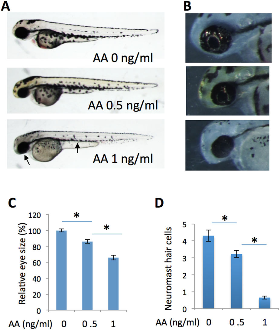
Fig. 7
Inhibition of energy ATP production by antimycin A partially phenocopies the prps1a;prps1b mutant phenotypes.
(A) Antimycin A treatment of wild-type embryos produces a dose-dependent reduction in melanocytes and eye size. Antimycin A concentrations are indicated. Arrows point to small eye and melanocyte reduction in the embryo treated with 1 ng/ml of antimycin A. (B) Dose-dependent reduction of retinal iridophores in the treated embryos. (C) Quantification of eye size reduction. Eye size is measured by Image J. A significant reduction is detected between antimycin A at 0 ng/ml and 0.5 ng/ml (t-test, p < 0.001), and between antimycin A at 0.5 ng/ml and 1 ng/ml (t-test, p = 0.019). (D) Dose-dependent reduction of neuromast hair cells in antimycin A-treated embryos. A significant reduction is detected between antimycin A at 0 ng/ml and 0.5 ng/ml (t-test, p = 0.008), and between antimycin A at 0.5 ng/ml and 1 ng/ml (t-test, p < 0.001). Eye area and neuromast hair cell analysis were performed with 10 embryos for each group. Graphs show the mean and s.e.m. Replication of the experiment produced similar results.