
IMAGE
Fig. 3
- ID
- ZDB-IMAGE-170628-2
- Genes
- Publication
- Plavicki et al., 2014 - Construction and characterization of a sox9b transgenic reporter line.
- All Figures
- Figures for Plavicki et al., 2014
Image
|
Figure Caption
Fig. 3
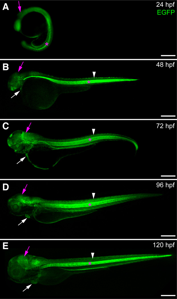
Expression of sox9b:EGFP during embryonic and larval development
(A–E) Lateral views of sox9b:EGFP embryos and larvae. (A) Epifluorescent image at 24 hpf. (B–E) Confocal images at 48 hpf (B), 72 hpf (C), 96 hpf (D) and 120 hpf (E). sox9b:EGFP expression is detected in the brain (purple arrows), eye, heart (white arrows), jaw, spinal cord (white arrowhead) and notocord (pink asterisks). Scale bars, 100 microns.
Figure Data
Acknowledgments
This image is the copyrighted work of the attributed author or publisher, and
ZFIN has permission only to display this image to its users.
Additional permissions should be obtained from the applicable author or publisher of the image.
Full text @ Int. J. Dev. Biol.